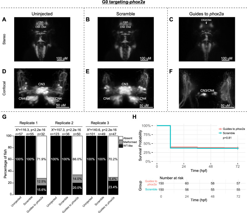
IMAGE

Figure 2.

ID
ZDB-IMAGE-250403-30
Source
Figures for Jurgens et al., 2025
Image
Figure Caption

Figure 2.

G0 zebrafish targeting of phox2a. Representative images obtained by stereomicroscope (AC) and confocal microscopy (DF) of uninjected (A, D), scramble-injected (B, E), or phox2a-targeting guide-injected G0 Tg(isl1:GFP) zebrafish at 72 hpf. Stereomicroscope images for each treatment group correspond to the confocal images obtained from the same fish in the panel below. Because residual motor neurons from CN3 and/or CN4 could not be definitively assigned to either of these specific motor neuron nuclei, CN3/CN4 were labeled as a single entity in Guide-targeted fish. Note apparent absence (C) or paucity (F) of CN3/CN4 motor neurons following phox2a G0 mosaic knockout. (G) Barplots showing the percentages of zebrafish exhibiting wild-type-like (“WT-like”), malformed, or absent CN3/CN4 motor nuclei, as scored under the dissecting stereomicroscope in G0 zebrafish at 72 hpf. Total numbers of fish in each group are given above the corresponding bar. Data are shown from three experimental replicates. Pearson's 3 × 3 χ2 test with 4 degrees of freedom; P values and χ2 values provided for each replicate. (H) Kaplan-Meier survival curves demonstrating the relative survival probabilities of scramble-injected (blue line) and guide-injected (pink line) zebrafish over the first 72 hours of life. “Number at risk” below the plot provides the counts of surviving embryos in each group taken every 24 hpf over a 72 hpf period. Relative survival probabilities of phox2a-targeting and scrambled gRNA-injected embryos were compared by the log-rank test. Displayed data were derived from a single experimental replicate, but measurements were taken for three experimental replicates, all of which showed the same trend.

Acknowledgments
This image is the copyrighted work of the attributed author or publisher, and ZFIN has permission only to display this image to its users. Additional permissions should be obtained from the applicable author or publisher of the image. Full text @ Invest. Ophthalmol. Vis. Sci.