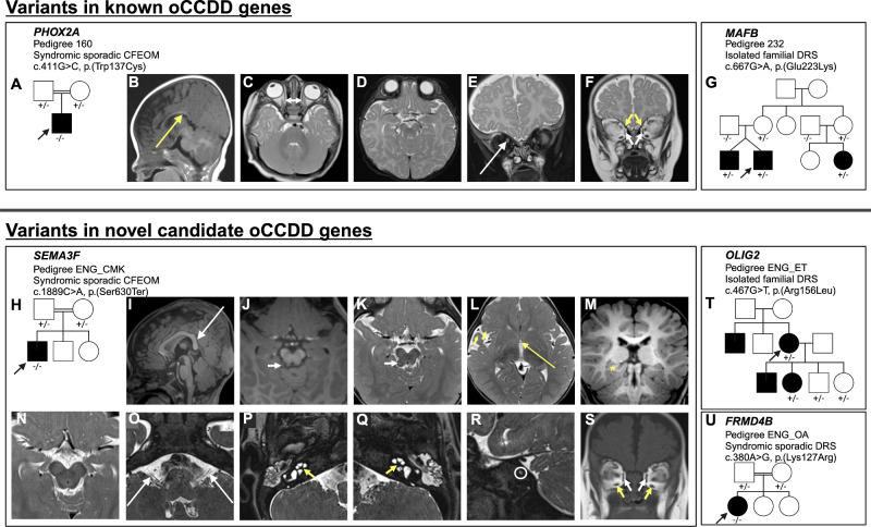
IMAGE

Figure 8.

ID
ZDB-IMAGE-250403-29
Source
Figures for Jurgens et al., 2025
Image
Figure Caption

Figure 8.

The oCCDD pedigrees with functionally validated candidate genes/variants and brain MR images of probands with homozygous PHOX2A and SEMA3F variants. (A, G, H, T, U) Schematics of pedigrees segregating variants of uncertain significance in known or novel candidate oCCDD genes. (BF) Images from a 1.5T Siemens brain MRI of the pedigree 160 proband at four months of age that has CFEOM and harbors a homozygous PHOX2A variant. (B) Sagittal T1 fluid–attenuated inversion recovery 4 mm–thick image reveals abnormal anatomy of the corpus callosum with a somewhat down-slanted posterior body and splenium (long yellow arrow). (C) Axial turbo spin echo (TSE) T2-weighted 4 mm–thick image shows diminutive medial rectus muscles (short double-headed white arrow). (D) Axial TSE T2 weighted 4 mm thick image at the level of the midbrain and interpeduncular cistern does not show oculomotor nerves. (E, F) Coronal TSE T2-weighted 4 mm–thick images with and without fat-suppression show asymmetric positioning of the optic nerves, higher on the right (long white arrow), diminutive medial rectus muscles (short white arrows), and small superior oblique muscles (short yellow arrows). Because of slice thickness and slice angle, the superior rectus muscles were not as readily assessed but also appeared slightly small. (IS) The ENG_CMK proband that has CFEOM and harbors a homozygous SEMA3F had MR imaging at 12 months of age obtained on a 3T Siemens Skyra (IM, OS) and three months of age obtained on a 1.5T Siemens unit (N) variant. I, Midline sagittal T1 magnetization prepared rapid gradient echo (MPRAGE) 1 mm–thick image demonstrates abnormal anatomy of the corpus callosum with a somewhat down-slanted posterior body and splenium (long white arrow). (J) Axial 1 mm reformatted MPRAGE and (K) 2.5 mm–thick TSE T2-weighted images show a small protuberance off or along the right side of the tectum (short white arrows) that appears hypointense on T1 and heterogeneous on T2 with central high-signal and peripheral low-signal intensity. This lesion is of uncertain etiology but was present in retrospect on an examination the previous year. (L) Axial 2.5–mm thick TSE T2-weighted image demonstrating a diminutive anterior commissure (long yellow arrow) and slight underdevelopment of the right frontal and temporal opercula (short yellow arrow). (M) Reformatted 1 mm coronal MPRAGE image shows asymmetry of the hippocampal formations and medial temporal lobes with the right side appearing mildly misshapen (yellow asterisk). N, Axial 2.5 mm–thick T2-weighted image at the level of the midbrain does not show oculomotor nerves at the level of the interpeduncular cistern; O, Axial 0.44 mm–thick T2 Sampling perfection with application optimized contrasts using different flip angle evolution (SPACE) image shows that the cisternal segments of the vestibulocochlear nerves are present but small (long white arrows) as they course posterior and parallel to the facial nerves. (P, Q) T2 SPACE images through the inner ears show dysmorphic, thickened cochlear modioli with stenotic cochlear apertures, more pronounced on the left (short yellow arrows). The apices of the cochleae appear mildly flattened. (R) Sagittal oblique T2 space MRI shows marked stenosis of the left internal auditory meatus (white circle) with only one cranial nerve visible instead of the expected four (facial, cochlear, and superior and inferior vestibular nerves). (S) Coronal 5 mm–thick T1-weighted image of the brain at the level of the orbits shows small medial rectus (short white arrows) and inferior rectus muscles (short yellow arrows). The superior rectus muscles are likely also small but are suboptimally assessed because of slice thickness.

Acknowledgments
This image is the copyrighted work of the attributed author or publisher, and ZFIN has permission only to display this image to its users. Additional permissions should be obtained from the applicable author or publisher of the image. Full text @ Invest. Ophthalmol. Vis. Sci.