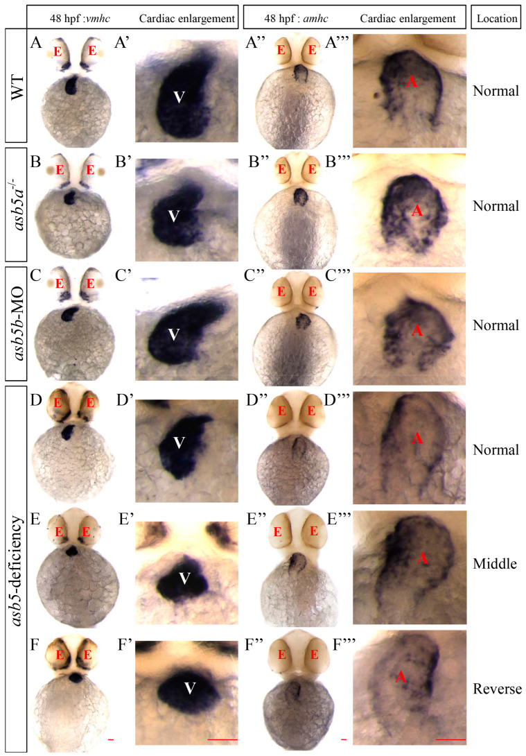
IMAGE

Figure 4

ID
ZDB-IMAGE-250329-88
Source
Figures for Zhou et al., 2025
Image
Figure Caption

Figure 4

WISH results using vmhc and amhc probes at 48 hpf in zebrafish embryos. (AF) Schematic representations of ventricular positions labeled by the vmhc probe across different groups at 48 hpf. (A′F′) Enlarged images of ventricular tissues labeled by the vmhc probe in these groups. (A″F″) Schematic representation of the atrial positions labeled by the amhc probe across various groups at 48 hpf. (A′′′F′′′) Enlarged images of atrial tissues labeled by the amhc probe in these groups. red E: eyes; white V: ventricle; red A: atrium; Normal: Normal position; Middle: Middle position; Reverse: Reverse position; Scale bar = 100 µm (belly up view).

Acknowledgments
This image is the copyrighted work of the attributed author or publisher, and ZFIN has permission only to display this image to its users. Additional permissions should be obtained from the applicable author or publisher of the image. Full text @ Int. J. Mol. Sci.