
|
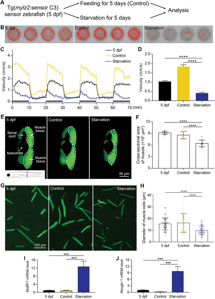
Figure 3
Establishment of a starvation‐induced muscle atrophy model. A) The muscle atrophy model is induced by starvation. B) The swimming trajectories of zebrafish for 5 min. C) Zebrafish locomotion tracking shows the decreased swimming velocity after starvation (n = 24 zebrafish for each group). D) The quantified results of zebrafish locomotion tracking (n = 24 zebrafish for each group). E) Images showing the cross‐sectional areas of muscle tissues. The imaging position and anatomical structures are indicated in the first image. Half of the muscle tissue is imaged, which is circled by a dashed line. F) The quantified results show that the cross‐sectional area of muscle tissues decreased after starvation (n = 10 zebrafish for each group). G) Images of dissociated muscle tissues show a decrease in the diameter of muscle cells after starvation‐induced muscle atrophy. H) The quantified results show that the diameter of muscle cells decreased after starvation (n = 60 cells for each group). I) The mRNA level of MuRF1 increased after starvation. J) The mRNA level of Atrogin‐1 increased after starvation. The size of each scale bar is indicated in each image. ***