IMAGE

Figure 1

ID
ZDB-IMAGE-250328-20
Source
Figures for Jia et al., 2025
Image
Figure Caption

Figure 1

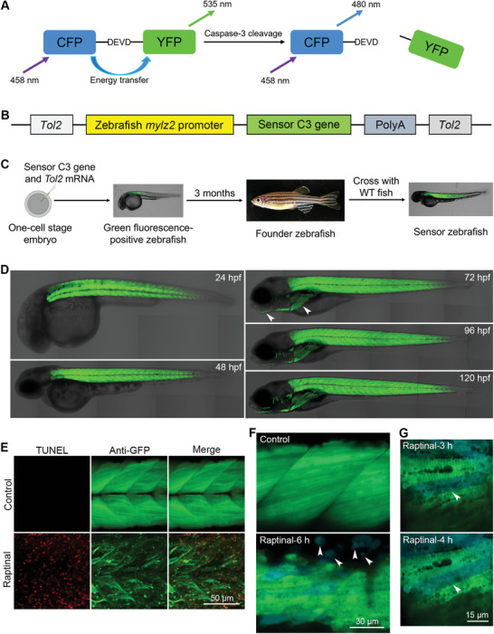
Generation of transgenic sensor zebrafish expressing an apoptotic biosensor in muscle cells. A) The structure and principle of sensor C3. B) The design and components of the Tol2‐based transgenic plasmid. C) The process of generating transgenic Tg(mylz2:sensor C3) zebrafish. D) Expression of sensor C3 in muscle cells of Tg(mylz2:sensor C3) zebrafish. The jaw and pectoral fin are indicated with arrowheads. The developmental time is indicated in each image. E) TUNEL assays showing the muscle cell apoptosis after the treatment of Raptinal for 8 h. F) FRET imaging showing the Raptinal‐induced muscle cell apoptosis in Tg(mylz2:sensor C3) zebrafish. The apoptotic bodies are indicated with arrowheads. G) Apoptotic muscle cells induced by Raptinal could maintain their morphology after caspase‐3 activation for more than 1 h. One blue muscle cell is indicated by an arrowhead to show the morphology. The size of each scale bar is indicated in each image.

Acknowledgments
This image is the copyrighted work of the attributed author or publisher, and ZFIN has permission only to display this image to its users. Additional permissions should be obtained from the applicable author or publisher of the image. Full text @ Adv Sci (Weinh)