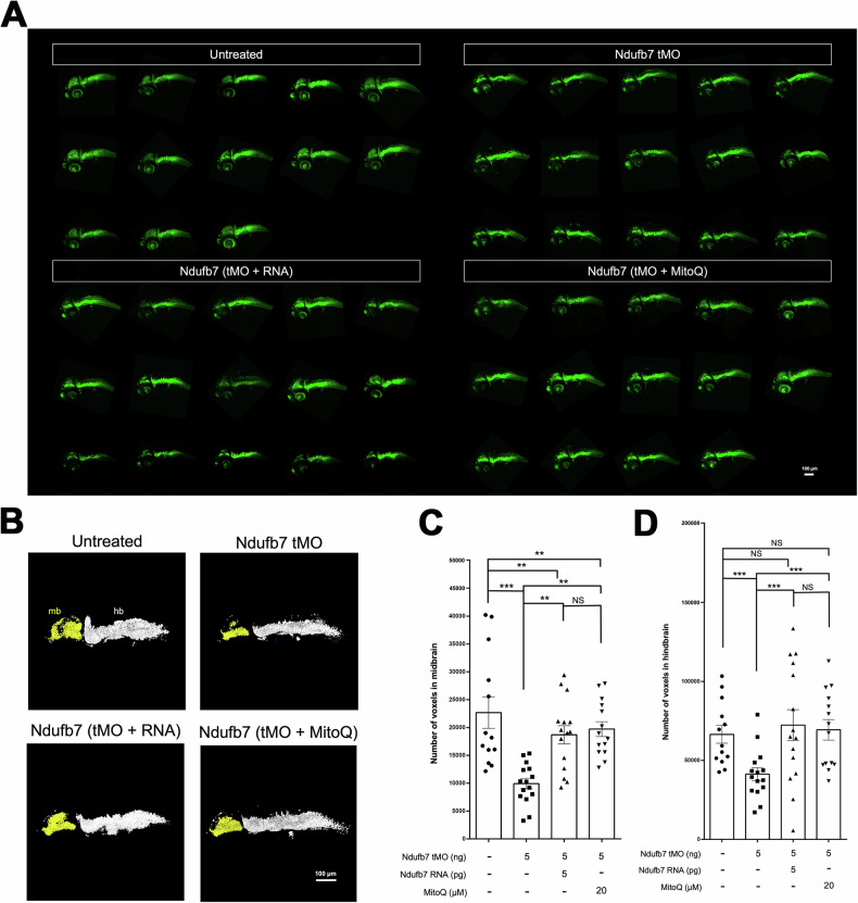
Fig. 5
Knockdown of Ndufb7 reduces the neuronal volume of the brain.
Transgenic Tg(Huc:kaede) zebrafish embryos were treated as described in Fig. 3, cultured until 48 hours post-fertilization, observed using a 10X objective, and photographed under a black field using a GFP cube under confocal microscopy. A All images are presented in lateral view. B The midbrain (mb) and hindbrain (hb) are pseudocolored in yellow and white, subjected to volume measurement using the Metamorph software, as shown in C midbrain and D hindbrain. The volume of each brain compartment was measured in voxels. Data from three independent experiments are presented, analyzed, and shown as described in Fig. 3. NS not significant; **p < 0.01; ***p < 0.001.
This image is the copyrighted work of the attributed author or publisher, and
ZFIN has permission only to display this image to its users.
Additional permissions should be obtained from the applicable author or publisher of the image.
Full text @ Cell Death Discov
Your Input Welcome
Thank you for submitting comments. Your input has been emailed to ZFIN curators who may contact you if
additional information is required.
Oops. Something went wrong. Please try again later.