IMAGE

Fig. 3

ID
ZDB-IMAGE-250304-3
Source
Figures for Chen et al., 2025
Image
Figure Caption

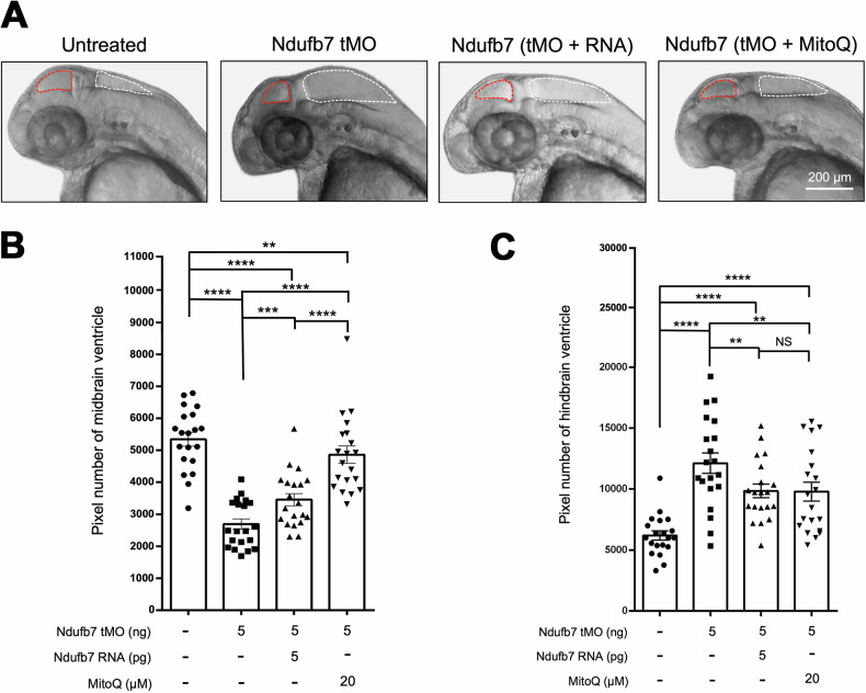
Fig. 3 Knockdown of Ndufb7 changes the sizes of brain ventricles.

We microinjected 1-cell stage zebrafish embryos without (untreated) or with indicated reagents and photographed them under a stereomicroscope at 48 hours post-fertilization. A Representative images for each treatment are presented. The ventricles are enclosed in red and white dotted lines for the midbrain and hindbrain, respectively. The area of the ventricle was measured in pixels using the Image J software. All data are shown in a scatter plot with mean ± standard error of the mean (SEM) for each treatment shown at the bottom of each column for B midbrain and C hindbrain, respectively, from three independent experiments. **p < 0.01; ***p < 0.001; ****p < 0.0001.

Figure Data
Acknowledgments
This image is the copyrighted work of the attributed author or publisher, and ZFIN has permission only to display this image to its users. Additional permissions should be obtained from the applicable author or publisher of the image. Full text @ Cell Death Discov