
Image
|
Figure Caption
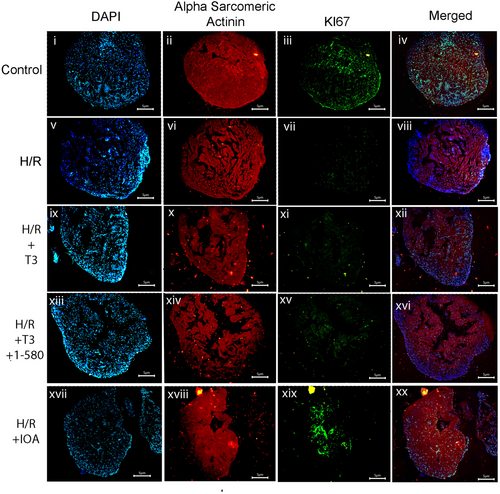
Fig. 9 Immunofluorescence study showing decreased nuclear expression of Ki67 in T3 + H/R or H/R alone compared to control in cardiac tissue sections. Tissue sections were incubated with primary antibodies against Ki67 (Parts iii, vii, xi, xv, xix) followed by Alexa fluor 488 secondary antibody. All the sections were counter stained with alpha sarcomeric actinin antibody (Parts iii, vii, xi) followed by Alexa fluor 633 secondary antibody (ii, vi, x, xiv, xviii) and nuclei with DAPI (Parts I, v, ix, xiii, xvii). Parts iv, viii, xii, xvi and xx show merged images (scale bar = 5 μm).
Acknowledgments
This image is the copyrighted work of the attributed author or publisher, and
ZFIN has permission only to display this image to its users.
Additional permissions should be obtained from the applicable author or publisher of the image.
Full text @ Wound Repair Regen.