IMAGE

Fig 6

ID
ZDB-IMAGE-250203-14
Source
Figures for Wang et al., 2024
Image
Figure Caption

Fig 6

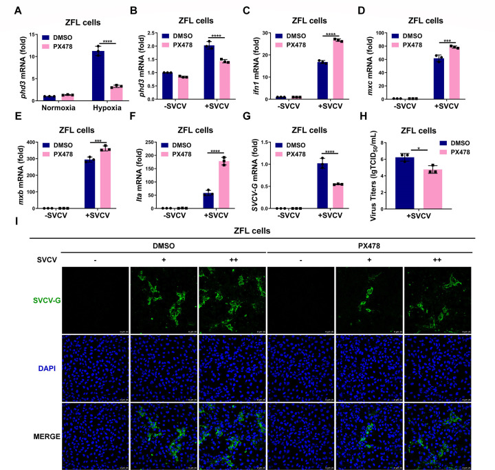
HIF-1α inhibitor PX478 enhances the antiviral ability against SVCV infection and inhibits SVCV replication in ZFL cells. (A) qPCR analysis of phd3 mRNA in ZFL cells treated with PX478 (10 µM) for 12 h, followed by hypoxia treatment for 24 h. (B) qPCR analysis of phd3 mRNA in ZFL cells treated with PX478 (10 µM) for 12 h, followed by infected with or without SVCV for 24 h. (C-F) qPCR analysis of ifn1 (C), mxc (D), mxb (E), and lta (F) mRNA in ZFL cells treated with PX478 (10 µM) for 12 h, followed by infected with or without SVCV for 24 h. (G) qPCR analysis of SVCV-G mRNA in ZFL cells treated with PX478 (10 µM) for 12 h, followed by infected with or without SVCV for 24 h. (H) ZFL cells were treated with or without PX478 (10 µM) for 12 h, followed by those infected with SVCV for 24 h. Culture supernatant was collected, and viral titers were measured by a 50% tissue culture–infective dose (TCID50) assay on EPC cells. (I) ZFL cells seeded on 35 mm glass bottom cell culture dishes were treated with PX478 (10 µM) for 12 h, followed by infected with increased gradient SVCV for 24 h. The cells were fixed and stained with anti-SVCV-G antibody and subsequent confocal microscopy analysis. Scale bar = 25 µm.

Acknowledgments
This image is the copyrighted work of the attributed author or publisher, and ZFIN has permission only to display this image to its users. Additional permissions should be obtained from the applicable author or publisher of the image. Full text @ J. Virol.