
|
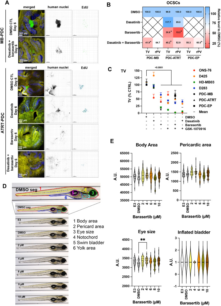
Fig. 6 Barasertib effectively blocks patient-derived cell growth in the tissue context as is not toxic. Confocal microscopy IFA of OCSC implanted MB-PDC and ATART-PDC 5 days after treatments with 500 nM Barasertib or Dasatinib or the combination of Barasertib and Dasatinib. Green: Anti-human nucleoli antibody (PDCs), red: Click-iT® EdU, blue: anti-Cabindin, yellow: anti-GFAP. MB: medulloblastoma, ATRT: atypical teratoid rhabdoid tumor, EP: ependymoma. Human nuclei are displayed in inverted greyscale and EdU incorporation as volume renderings from Z-stacks. B) Heat map of TV and rPV of PDC-OCSC co-cultures after 5-days of treatments (n≥3 biological replicas, *p 0.05). C-D) TV and the rPV of all cell lines and PDCs used (Mean + SEM). C) Compilation of all TVs from the OCSC experiments. D) Representative VAST images of lateral view of zebrafish in the different treatment groups. The first image corresponds to a control animal with segmentation of anatomical regions studied: body area (red), eye size (green), pericardiac area (light blue), inflated bladder (pink), yolk area (blue), and notochord (yellow). E) Quantification of a subset of morphological areas shown in D.