
|
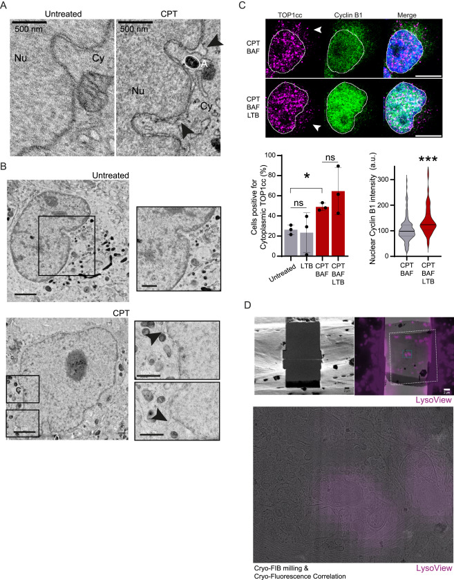
Fig. S5 CPT induces nuclear envelope blistering and TOP1cc exit to the cytoplasm, related to Figure 5 (A) Representative electron microscopy images for a distinct set of cells. Treatment was conducted for 30 min with 50 nM CPT. Scale bar, 500 nm. Arrows pointing at blister structures. Nu, nucleoplasm; Cy, cytoplasm; A, autophagosome. (B) Representative electron tomographic slices fixed by high-pressure freezing showing nuclear envelope blister in HeLa. Treatment was conducted for 30 min with 50 nM CPT. Scale bar, 2 μm (left); scale bar, 1 μm (zoom, right). Arrows pointing at blister structures. (C) Immunofluorescence of TOP1cc foci and cyclin B1 after 3 h of treatment with 50 nM CPT (n = 3). Cyclin B1 is used as a control for the inhibition of exportin by leptomycin B (LTB). Scale bar, 10 μm. (Left) Quantification of positive cells for cytoplasmic TOP1cc foci. One-way ANOVA. (Right) Cyclin B1 intensity per nuclei (quantification for a representative experiment). t test. Error bar, SD. ∗p < 0.05; ∗∗∗p < 0.0005; ns, not significant. (D) Cryo-focused ion beam (FIB) milling and cryo-fluorescence correlation images of cells after 30 min of 50 nM CPT treatment. Lysosomes are stained with LysoTracker. (Top) Ion beam image of a final lamellae after targeted milling and correlation between the SEM image of a lamellae, the transmission electron microscopy (TEM) medium magnification montage of the same lamellae acquired at 15,000× (white dashed lines) and the fluorescent molecularly imprinted polymer (MIP) acquired post milling. (Bottom) Close-up view on the TEM medium magnification correlated with LysoTracker fluorescence used for targeted acquisition of the tilt series. Scale bar, 2 μm.
Reprinted from Cell, 187(20), Lascaux, P., Hoslett, G., Tribble, S., Trugenberger, C., Antičević, I., Otten, C., Torrecilla, I., Koukouravas, S., Zhao, Y., Yang, H., Aljarbou, F., Ruggiano, A., Song, W., Peron, C., Deangeli, G., Domingo, E., Bancroft, J., Carrique, L., Johnson, E., Vendrell, I., Fischer, R., Ng, A.W.T., Ngeow, J., D'Angiolella, V., Raimundo, N., Maughan, T., Popović, M., Milošević, I., Ramadan, K., TEX264 drives selective autophagy of DNA lesions to promote DNA repair and cell survival, 5698-5718.e26, Copyright (2024) with permission from Elsevier. Full text @ Cell