
|
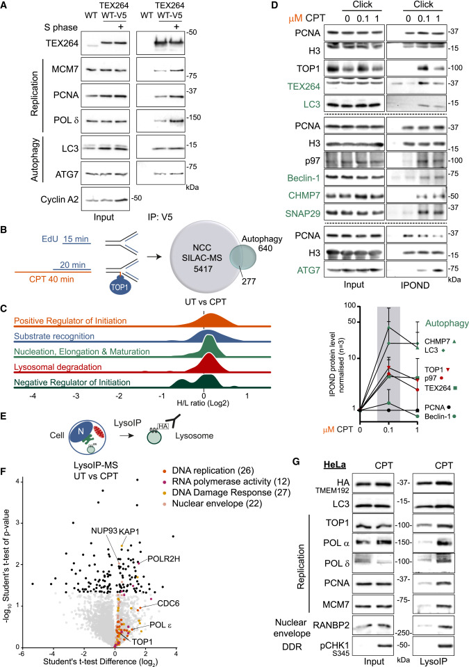
Fig. 1 Crosstalk between autophagy and DNA replication upon replication fork stalling by CPT (A) Co-immunoprecipitation of TEX264-WT-V5 in HeLa TEX264KO background in asynchronized or S-phase-synchronized cells (n = 3). (B) Strategy for replisome proteomics analysis. Venn diagram of the replisome found by nascent chromatin capture (NCC) coupled to stable isotope labeling by amino acids in cell culture (SILAC) and mass spectrometry (MS) analysis, NCC-SILAC-MS63 overlapped with all known autophagy factors. (C) Ridge regression plot of known autophagy factors at replication fork identified by SILAC-NCC-MS.63 (D) iPOND performed after 40 min of 100 nM CPT (0.1 μM) or 1 μM CPT. Representative blots from different biological repeats and quantification (n = 3). Error bar, SD. (E) Strategy for purification of intact lysosomes; LysoIP. (F) Proteome profiling of lysosomes purified by LysoIP after 6 h of 50 nM CPT. Proteins differentially expressed (−10log p >1.301) shown with full dark circles. (G) LysoIP performed in S-phase-synchronized HeLa after 6 h of 50 nM CPT. See also Figure S1 and Tables S1 and S2.
Reprinted from Cell, 187(20), Lascaux, P., Hoslett, G., Tribble, S., Trugenberger, C., Antičević, I., Otten, C., Torrecilla, I., Koukouravas, S., Zhao, Y., Yang, H., Aljarbou, F., Ruggiano, A., Song, W., Peron, C., Deangeli, G., Domingo, E., Bancroft, J., Carrique, L., Johnson, E., Vendrell, I., Fischer, R., Ng, A.W.T., Ngeow, J., D'Angiolella, V., Raimundo, N., Maughan, T., Popović, M., Milošević, I., Ramadan, K., TEX264 drives selective autophagy of DNA lesions to promote DNA repair and cell survival, 5698-5718.e26, Copyright (2024) with permission from Elsevier. Full text @ Cell