
|
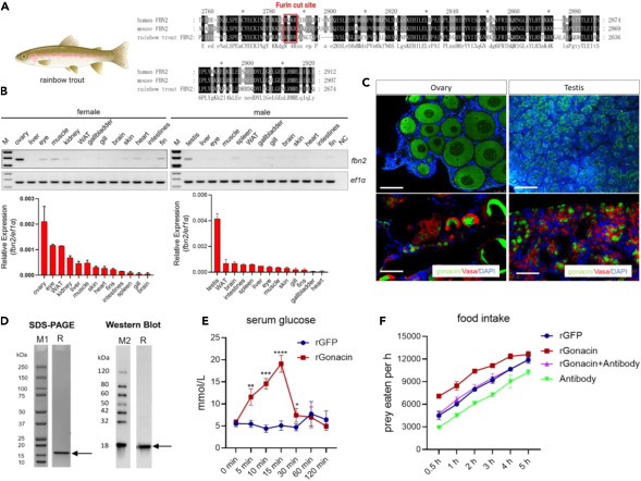
Fig. 8 Identification of gonacin in rainbow trout exhibiting gonadal expression and showing glucogenic and orexigenic activities (A) Identification of gonacin in the C-terminal end of rainbow trout Fbn2 with a canonical furin cleavage position. (B) Expression of rainbow trout fbn2 mRNA in different adult tissues detected by RT-PCR followed by electrophoresis (upper panel) and real-time PCR (lower panel). (C) Expression of rainbow trout gonacin in ovary and testis detected by immunostaining. Localization of gonacin signal (Green, upper panel) and co-localization of gonacin (Green) and Vasa (Red) signal (lower panel) in ovary and testis of rainbow trout at 8 months post-fertilization. (D) Characterization of recombinant rainbow trout gonacin protein (rGonacin). Coomassie blue staining and immunoblotting of bacteria-derived rGonacin. M1, M2: marker proteins; R, rGonacin. (E) Serum glucose levels were measured after an intraperitoneal injection of rGonacin in vivo. ∗, p < 0.05; ∗∗, p < 0.01; ∗∗∗, p < 0.001; ∗∗∗∗, p < 0.0001, compared with control (n = 7 in each group). two-way ANOVA with Bonferroni post-test was used to calculate the p value. (F) Food intake was measured at indicated time points after injection of a single dose of rGFP, 5 μg rGonacin or 4 μL anti-gonacin antibodies or following co-injection of 5 μg rGonacin and 4 μL anti-gonacin.