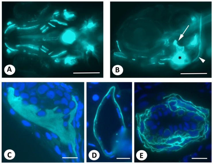
Live staining of early postembryonic zebrafish embryos with DAF-FM DA. (A,B). Live staining with DAF-FM DA, according to published protocols, reveals signals in bony structures (opercular, arrow; cleithrum, arrowhead), as well as in the heart (asterisk) and around the notochord, which is consistent with earlier reports. Axio Observer image. (C–E). Three µm GMA sections of the specimen shown in (A,B), counterstained with DAPI, showing the opercular bone (C), notochord (D), and bulbus arteriosus (E). Note that nuclei lie outside fluorescent domains. Scale bars in (A,B) = 200 µm, in (C,E) = 20 µm, in (D) = 10 µm.
Acknowledgments
This image is the copyrighted work of the attributed author or publisher, and
ZFIN has permission only to display this image to its users.
Additional permissions should be obtained from the applicable author or publisher of the image.
Full text @ Biomolecules
Your Input Welcome
Thank you for submitting comments. Your input has been emailed to ZFIN curators who may contact you if
additional information is required.
Oops. Something went wrong. Please try again later.