
|
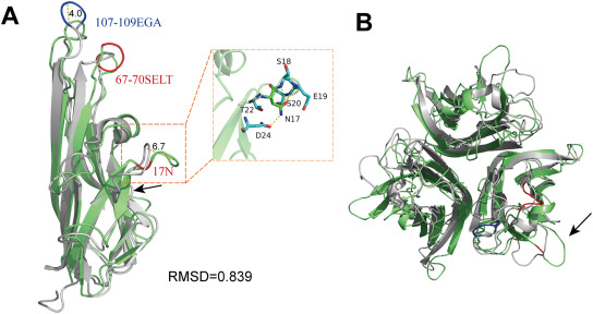
Fig. 5 Structural superposition of zTNF-α1 and hTNF-α. (A) Structural superposition of zTNF-α1 and hTNF-α monomers. zTNF-α1 is shown in green. hTNF-α is shown in light gray. The black arrow indicates the additional β-strand in zTNF-α1. hTNF-α has three amino acid insertions at positions 107–109, which are indicated in blue. The amino acid insertions at positions 17 and 67–70 of zTNF-α1 are shown in red, and the corresponding distances are indicated by yellow dashed lines and labeled with the length in angstroms. The insert on the right shows the key residues interacting with 17N resulting in hydrophobic oligomerization, which are shown as sticks. Hydrogen bonds are indicated with a yellow dashed line. (B) Top view of the structural alignment of the zTNF-α1 trimer and the hTNF-α trimer. The black arrow indicates the region folding outward resulting from 17N.