
|
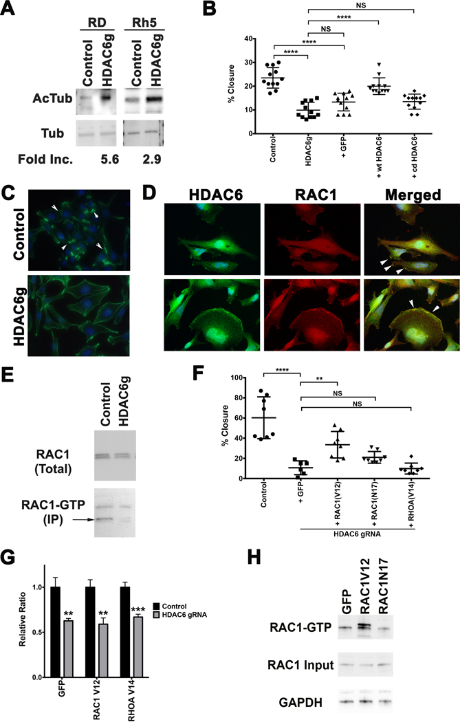
Figure 4. (A) Western blots against acetylated alpha-tubulin and alpha-tubulin in RD and Rh 5 cells with tamoxifen-induced CRISPR/Cas9-mediated targeted disruption of HDAC6 (day 6 post-tamoxifen). Fold increase in the levels of acetylated alpha-tubulin following normalization to the levels of alpha-tubulin was quantified using Image J. (B) Scratch assay assessing effects of adding back Cas9-resistant wild-type (wt) HDAC6 and catalytically-dead (cd) HDAC6 in RD cells with tamoxifen-induced HDAC6 targeting. Results at 16 hours post-scratch are shown from one representative experiment of at least 3 repeats. (C) Phalloidin staining in RD cells with no tamoxifen control and tamoxifen-induced HDAC6 CRISPR targeting following serum starvation and 15-minute EGF (50 ng/mL) treatment at day 6 post-tamoxifen treatment. Arrowheads point to representative areas of membrane ruffles and filopodia formation. Green = phalloidin, Blue = DAPI. (D) Double IF against HDAC6 (green) and RAC1 (red) in RD, showing HDAC6 and RAC1 expression in the regions of membrane ruffles (top panels) and folds (bottom panels), also highlighted by the arrowheads in merged panels. (E) RAC1 GTP pulldown assay in RD cells harboring no tamoxifen control and tamoxifen-induced HDAC6 CRISPR targeting. (F) Summary of scratch assays assessing the effects of lentiviral overexpression of GFP as a control, RAC1V12, RAC1N17 and RHOAV14 in the presence of tamoxifen-induced CRISPR-mediated targeted disruption of HDAC6 in RD cells at 16 hours post-scratch following 24 hours of serum starvation and 15 minutes of EGF (50 ng/mL) treatment. (G) Summary of cell growth change by cell counts over 6 days assessing the effects of overexpressing GFP as a control, RAC1V12 and RAC1N17 on cell growth of RD cells with tamoxifen-induced targeted disruption of HDAC6. Results were normalized to the no tamoxifen control for each comparison and represent the average of 4 replicates in one of 3 independent experiments. (H) RAC1-GTP pulldown assay in RD cells overexpressing GFP, RAC1V12 and RAC1N17 following 24 hours of serum starvation and 15 minutes of EGF (50 ng/mL) treatment. Each error bar in B, F and G represents standard deviation. Two-tailed t-test in B and G, one-way ANOVA test in F; NS = no significance, p < 0.05; ** = p < 0.01; *** = p < 0.001; **** = p < 0.0001.