
Image
|
Figure Caption
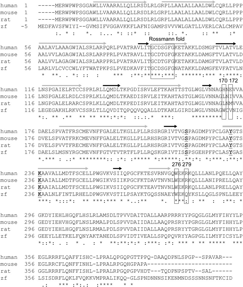
Fig. 2 Alignment of human, mouse, rat and zebrafish 11β-HSD2 protein sequences. Symbols are coded as followed: grey arrow, α-helix; black arrow, β-sheets; rectangles, cofactor-binding site in the Rossmann-fold, and positions 170, 172, 276 and 279; underlined and bold letter, active site; (*), fully conserved residues; (:), residues with considerably similar properties; (.), residues with moderately similar properties.
Acknowledgments
This image is the copyrighted work of the attributed author or publisher, and
ZFIN has permission only to display this image to its users.
Additional permissions should be obtained from the applicable author or publisher of the image.
Full text @ Tox. App. Pharmacol.