
Fig. 9
- ID
- ZDB-IMAGE-130304-39
- Genes
- Publication
- Di Donato et al., 2013 - Characterization of the calcium binding protein family in zebrafish
- All Figures
- Figures for Di Donato et al., 2013
|
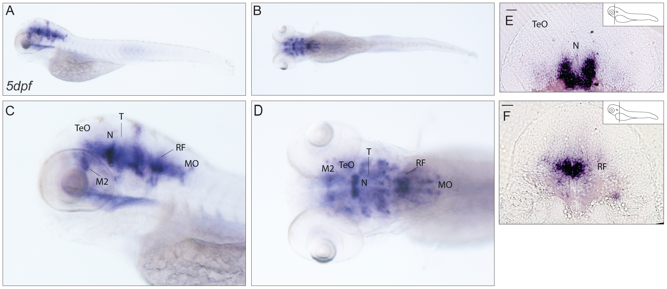
Fig. 9 Expression pattern of cabp7b.
(A–D) In situ localisation of cabp7b transcript in 5dpf larvae. Lateral (A,C) and dorsal views (B, D). A wide distribution of cabp7b mRNA in the brain was observed. (E,F) Transverse sections. Strongly stained cells are present in the midbrain, probably in the region of the nucleus of medial longitudinal fascicle (E) and in the hindbrain area anterior to the medulla oblongata most likely at the level of the reticular formation(F). Scale bar: 50 µm. M2: migrated posterior tubercular area; MO: medulla oblongata; N: nucleus of medial longitudinal fascicle; RF: reticular formation; T: midbrain tegmentum; TeO: tectum opticum.