FIGURE

FIGURE 3

ID
ZDB-FIG-250306-19
Publication
Liu et al., 2024 - Expression analysis of genes including Zfhx4 in mice and zebrafish reveals a temporospatial conserved molecular basis underlying craniofacial development
Other Figures
All Figure Page
Back to All Figure Page
FIGURE 3

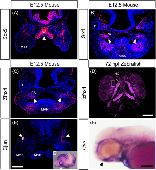
Protein localization in the developing head of mice and zebrafish. (A–D) Immunohistochemistry of selected transcription factors involved in chondrogenesis and osteogenesis using frontal sections of E12.5 mice head (A–C) and ventral view of 72 hours postfertilization (hpf) zebrafish larva (D). Samples are shown at the top, and proteins are shown on the left. White arrows indicate the protein detected in the nasal septum (A) and palate (B,C). (D) Immunofluorescence staining with anti‐Zfhx4 antibody in zebrafish embryos at 72 hpf. The developing ethmoid plate (zebrafish palate, dashed line) and Meckel's cartilage (mandibular, blacked) were stained with the antibody. Zfhx4 was also detected in the eye, ceratobranchial, ceratohyal, and hyosymplectic. (E,F) Similar expression domains of Cjun in both species were revealed using immunohistochemistry in the frontal section of E12.5 mice head, (E) cjun mRNA in the lateral view of E12.5 mice head, (E′) and the lateral view of 72 hpf zebrafish (F). Arrowheads indicate the expression surrounding the ocular region. E, eye; ep, ethmoid plate; MAN, mandibular; MAX, maxillary; m, Meckel's cartilage; NS, nasal septum; PS, palatal shelf; T: tongue. Scale bars: 500 μm in (E); 100 μm in (D,F).

Expression Data
Gene:
Antibody:
Fish:
Anatomical Terms:
Stage: Protruding-mouth

Expression Detail
Antibody Labeling
Phenotype Data

Phenotype Detail
Acknowledgments
This image is the copyrighted work of the attributed author or publisher, and ZFIN has permission only to display this image to its users. Additional permissions should be obtained from the applicable author or publisher of the image. Full text @ Dev. Dyn.