FIGURE

Figure 1

ID
ZDB-FIG-250127-188
Publication
Ren et al., 2024 - Knockout of dhx38 Causes Inner Ear Developmental Defects in Zebrafish
Other Figures
All Figure Page
Back to All Figure Page
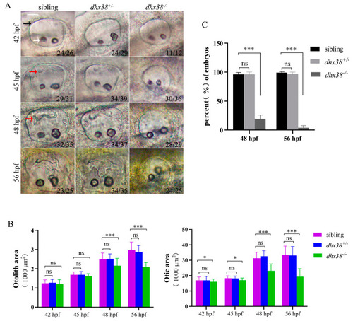
Figure 1

The deletion of dhx38 caused inner ear development defects: (A) The different morphological specificities of the inner ears of different genotypes at distinct developmental stages. Scale bar: 40 µm. The black arrow indicates the otic vesicle, the white arrow indicates the otolith, and the red arrow indicates semicircular canal protrusion. (B) Statistical analysis of the otic area and otolith area in the embryos of different genotypes at 42 hpf, 45 hpf, 48 hpf, and 56 hpf. Individual embryos were picked casually from particular types for statistical analysis. The otic and otolith areas were measured using SPOT Advanced software (version 4.6) in the focal plane exhibiting the maximal area [16]. n, the number of checked embryos. n = 40. (C) The percentages of embryos that had semicircular canal protrusions in the sibling, the dhx38+/ and dhx38/ groups at 48 hpf and 56 hpf. The data are presented as the mean ± SD. ns, p > 0.05; *, p < 0.05; ***, p < 0.001.

Expression Data

Expression Detail
Antibody Labeling
Phenotype Data
Fish:
Observed In:
Stage Range: High-pec to Long-pec

Phenotype Detail
Acknowledgments
This image is the copyrighted work of the attributed author or publisher, and ZFIN has permission only to display this image to its users. Additional permissions should be obtained from the applicable author or publisher of the image. Full text @ Biomedicines