
Fig. 5
- ID
- ZDB-FIG-250110-17
- Publication
- Smaili et al., 2024 - R391 human dominant mutation does not affect TubB4b localization and sensory hair cells structure in zebrafish inner ear and lateral line
- Other Figures
- All Figure Page
- Back to All Figure Page
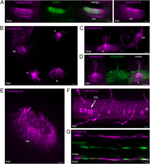
Expression of TubB4b[Chr5] in various tissues of zebrafish larva. A: Utricule (anterior macula: am) from double transgenic Tg(myo7aa:EGFP)+/0; Tg(tubB4b[Chr5]:TubB4b[Chr5]-mCherry)+/0 larva observed at 78 hpf (left panel) and anterior macula (am) from Tg(tubB4b[Chr5]:TubB4b[Chr5]-mCherry)+/0 larva imaged at 5 dpf (right panel). B: inner ear anterior half of fixed Tg(tubB4b[Chr5]:TubB4b[Chr5]-mCherry)+/0 larva displaying anterior crista (ac), lateral crista (lc) and 2 head neuromast (n). C: live imaging of lateral crista (lc) and posterior crista (pc) in a 72 hpf Tg(tubB4b[Chr5]:TubB4b[Chr5]-mCherry)+/0 larva. D: live imaging of a head neuromast (n) from a double transgenic Tg(tubB4b[Chr5]:TubB4b[Chr5]-mCherry)+/0; Tg(actb2:Mmu.Arl13b-GFP)+/0 larva. E: live imaging of olfactory epithelium (oe) in a 5 dpf Tg(tubB4b[Chr5]:TubB4b[Chr5]-mCherry)+/0 larva. F: live imaging of spinal chord (sc) and motoneurons (mn) in a 5 dpf Tg(tubB4b[Chr5]:TubB4b[Chr5]-mCherry)+/0 larva. G: live imaging of pronephros (p) in a double transgenic Tg(tubB4b[Chr5]:TubB4b[Chr5]-mCherry)+/0; Tg(actb2:Mmu.Arl13b-GFP)+/0 52 hpf larva. TubB4b[Chr5] indicates Tg(tubB4b[Chr5]:TubB4b[Chr5]-mCherry) fluorescence signal (A–G); EGFP indicates Tg(myo7aa:EGFP) fluorescence signal (A); Arl13b-EGFP indicates Tg(actb2:Mmu.Arl13b-GFP) fluorescence signal (D,G). Projection of various size images stacks. Scale bar = 10 μm for all images. |
Reprinted from Developmental Biology, 517, Smaili, W., Pezet, C., Marlin, S., Ernest, S., R391 human dominant mutation does not affect TubB4b localization and sensory hair cells structure in zebrafish inner ear and lateral line, 301-316, Copyright (2024) with permission from Elsevier. Full text @ Dev. Biol.