|
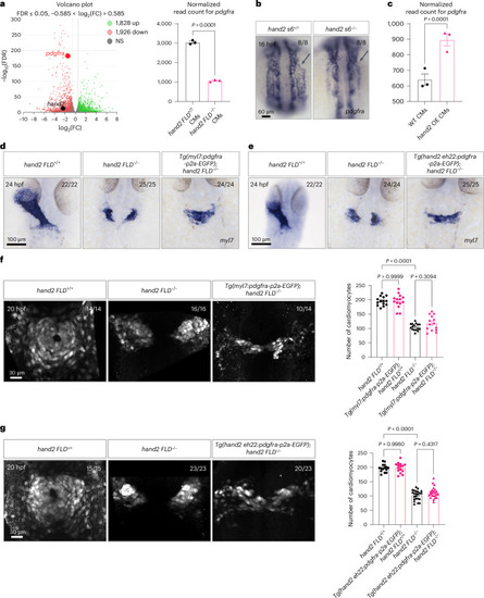
Hand2 regulates pdgfra expression to promote cardiac fusion. a, Left, transcriptomic analysis was performed using RNA extracted from sorted Tg(myl7:EGFP)+ cells from 20 hpf hand2 FLD+/? and hand2 FLD−/− sibling embryos. Right, normalized read count for pdgfra in 20 hpf hand2 FLD+/? and hand2 FLD−/− sibling Tg(myl7:EGFP)+ cells; error bars are mean ± s.e.m.; n = 3 biologically independent samples. b, In situ hybridization showing pdgfra expression in the anterior LPM (arrows) of 16 hpf hand2 s6+/? and hand2 s6−/− sibling embryos. c, Normalized read count for pdgfra in 20 hpf hand2 OE and WT sibling Tg(myl7:EGFP)+ cardiomyocytes; error bars are mean ± s.e.m.; n = 3 biologically independent samples. d, In situ hybridization showing myl7 expression in 24 hpf hand2 FLD+/+, hand2 FLD−/− and Tg(myl7:pdgfra-p2a-EGFP); hand2 FLD−/− sibling embryos. e, In situ hybridization showing myl7 expression in 24 hpf hand2 FLD+/+, hand2 FLD−/− and Tg(hand2 eh22:pdgfra-p2a-EGFP); hand2 FLD−/− sibling embryos. f, Left, Tg(myl7:EGFP)+ cells in 20 hpf hand2 FLD+/+, hand2 FLD−/− and Tg(myl7:pdgfra-p2a-EGFP); hand2 FLD−/− sibling embryos (of the 14 Tg(myl7:pdgfra-p2a-EGFP); hand2 FLD−/− embryos, 10 displayed a comparable number of myl7:EGFP+ cells as hand2 FLD−/− embryos and 4 displayed a few more myl7:EGFP+ cells). Right, quantification of cardiomyocyte numbers in 20 hpf hand2 FLD+/+ (n = 14), hand2 FLD−/− (n = 16), Tg(myl7:pdgfra-p2a-EGFP); hand2 FLD+/+ (n = 16) and Tg(myl7:pdgfra-p2a-EGFP); hand2 FLD−/− (n = 14) sibling embryos; error bars are mean ± s.e.m. g, Left, Tg(myl7:EGFP)+ cells in 20 hpf hand2 FLD+/+, hand2 FLD−/− and Tg(hand2 eh22:pdgfra-p2a-EGFP); hand2 FLD−/− sibling embryos (of the 23 Tg(hand2 eh22:pdgfra-p2a-EGFP); hand2 FLD−/− embryos, 20 displayed a comparable number of myl7:EGFP+ cells as hand2 FLD−/− embryos and 3 displayed a few more myl7:EGFP+ cells). Right, quantification of cardiomyocyte numbers in 20 hpf hand2 FLD+/+ (n = 15), hand2 FLD−/− (n = 23), Tg(hand2 eh22:pdgfra-p2a-EGFP); hand2 FLD+/+ (n = 23) and Tg(hand2 eh22:pdgfra-p2a-EGFP); hand2 FLD−/− (n = 23) sibling embryos; error bars are mean ± s.e.m. P values were calculated using an unpaired Student’s t test (a (right), c) or a one-way analysis of variance (ANOVA) multiple-comparison test (f (right), g (right)). All embryos are shown in dorsal views, anterior to the top. The proportion of embryos matching the image shown is indicated in the top right corner of each image. The scale bars apply to all images. FDR, false discovery rate; FC, fold change; CMs, cardiomyocytes; NS, not significant (P > 0.05).
|