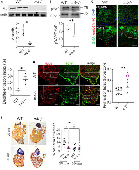
Loss of Mb promotes cardiomyocyte proliferation and regeneration in vivo (A) Western blot and quantification of Mb in WT and mb−/− zebrafish hearts; N = 3 biological replicates. (B) Representative western blot and quantification of pYAP and T-YAP WT and mb−/− zebrafish hearts; N = 3. (C) Zebrafish heart section demonstrating the staining for de-differentiation marker embCMHC in red and α-sarcomeric actin in green at 7 days post amputation, and quantification of embCMHC cardiomyocytes (Differentiation index: % of cardiomyocytes expressing embCMHC in border zone of injury) from WT and mb−/− hearts; N = 3 from each group. Scale bar: 100 μm. (D) Heart sections from WT and mb−/− zebrafish stained for Mef2c transcription factor (red) to identify cardiomyocytes and PCNA (green) to identify proliferating cells at 7 days post amputation: N = 7–9 individual zebrafish per group. Scale bar: 100 μm. (E) Representative histological sections of AFOG stained tissue from WT and mb−/− zebrafish at 20- and 30-days post amputation stained to visualize fibrin (red), collagen (blue) and cardiac muscle (orange). Graph demonstrates the quantification of scar area on each section normalized to total ventricular area; N = 9–16 individual zebrafish per group. Scale bar: 100 μm. Data are mean ± SEM. ∗p < 0.05, ∗∗p < 0.01, ∗∗∗∗p < 0.0001.
|
