FIGURE

Figure 2

ID
ZDB-FIG-230531-21
Publication
Zhao et al., 2023 - The manipulation of cell suspensions from zebrafish intestinal mucosa contributes to understanding enteritis
Other Figures
All Figure Page
Back to All Figure Page
Figure 2

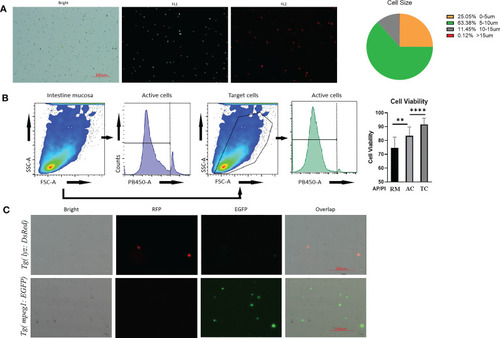
Quality control of prepared intestinal single cell suspension from Tg(lyz:DsRED2);Tg(mpeg1:EGFP). (A) Imaging by automated cell counter, cell under brightfield and fluorescence channels (FL1: 488nm; FL2:558nm). Scale bar: 100μm. The cellular diameter of the prepared intestinal mucosal cell suspension was calculated besides. (B) Cell viability reflected AO/PI staining during cytometric analysis. The stained cell number was counted by automatic cell counter. After AO/PI staining, 75% cell obtained from robbing method (RM) was alive, meanwhile for the blowing method, 80% of all cells (AC) and 90% of genetically fluorescence labeled immune cells, which were the target cells (TC), were alive for flow cytometric analysis. (C) Images of intestinal mucosal cells from Tg(lyz:DsRED2);Tg(mpeg1:EGFP). In single cell suspensions, DsRed labeled lyz+ cell and EGFP labeled mpeg1+ cells could be clearly observed, together with other unstained cells (shown in the brightfield). ** represented p < 0.01, **** represented p < 0.0001.

Expression Data

Expression Detail
Antibody Labeling
Phenotype Data

Phenotype Detail
Acknowledgments
This image is the copyrighted work of the attributed author or publisher, and ZFIN has permission only to display this image to its users. Additional permissions should be obtained from the applicable author or publisher of the image. Full text @ Front Immunol