|
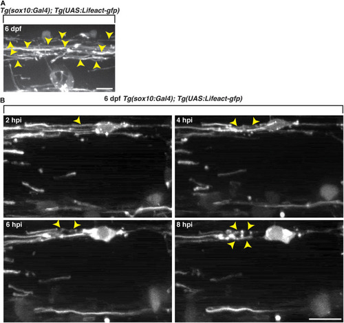
Oligodendrocyte cytoskeletal components dynamically change following exposure to lysolecithin. All images are lateral views of the spinal cord with anterior to the left and dorsal to the top. (A)In vivo imaging of Tg(sox10:Gal4; UAS:Lifeact-gfp) larvae at 6 dpf. Arrowheads denote the actin in sheath formation. (B) Following injection of lysolecithin into a 6 dpf Tg(sox10:Gal4; UAS-Lifeact-gfp) zebrafish, in vivo time-lapse imaging reveals that GFP+ F-actin dynamically changes from a sheath-like arrangement (2 hpi) to forming ovoid-like structures (8 hpi). Arrowheads identify the GFP+ F-actin changes in a sox10+ cell. Scale bars, 25 μm.
|