
FIGURE
Fig. 8
- ID
- ZDB-FIG-200325-202
- Publication
- Bagwell et al., 2020 - Notochord vacuoles absorb compressive bone growth during zebrafish spine formation
- Other Figures
- All Figure Page
- Back to All Figure Page
Fig. 8
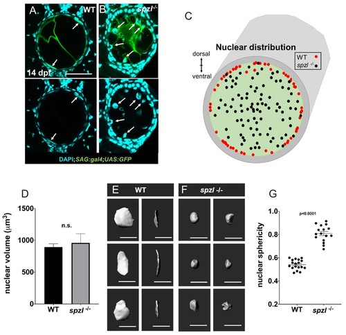
Nuclear distribution and shape indicate that hydrostatic pressure of the notochord is decreased in spzl mutants.( A–B) Confocal images of cross sections of 14 dpf WT and spzl-/- larvae section. Vacuolated cells are labeled with SAG:gal4;UAS:GFP and nuclei are stained with DAPI. Scale bar = 50 µm ( C) Plot of vacuolated cell nuclear distribution for WT (red, n = 29) and spzl-/- (black, n = 32). ( D) Nuclear volume in WT and spzl-/- at 14 dpf. ( E–F) Reconstructions of nuclei in WT ( E) and spzl-/- ( F) and two different viewing angles. Scale bar = 20 µm ( G) Sphericity of WT and spzl-/- nuclei at 14 dpf. p-values were determined by an un-paired t-test using Welch’s correction. |
Expression Data
| Gene: | |
|---|---|
| Fish: | |
| Anatomical Term: | |
| Stage: | Days 14-20 |
Expression Detail
Antibody Labeling
Phenotype Data
| Fish: | |
|---|---|
| Observed In: | |
| Stage: | Days 14-20 |
Phenotype Detail
Acknowledgments
This image is the copyrighted work of the attributed author or publisher, and
ZFIN has permission only to display this image to its users.
Additional permissions should be obtained from the applicable author or publisher of the image.
Full text @ Elife