
FIGURE
Fig. 6
- ID
- ZDB-FIG-191230-848
- Publication
- Kleinhans et al., 2019 - Standardized mounting method of (zebrafish) embryos using a 3D-printed stamp for high-content, semi-automated confocal imaging
- Other Figures
- All Figure Page
- Back to All Figure Page
Fig. 6
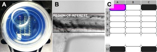
Imaging setup. |
Expression Data
Expression Detail
Antibody Labeling
Phenotype Data
Phenotype Detail
Acknowledgments
This image is the copyrighted work of the attributed author or publisher, and
ZFIN has permission only to display this image to its users.
Additional permissions should be obtained from the applicable author or publisher of the image.
Full text @ BMC Biotechnol.