
FIGURE
Fig. 4
- ID
- ZDB-FIG-171227-37
- Publication
- Mei et al., 2014 - Functional characterization of Prickle2 and BBS7 identify overlapping phenotypes yet distinct mechanisms
- Other Figures
- All Figure Page
- Back to All Figure Page
Fig. 4
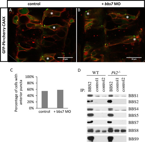
Normal Pk localization in bbs7 knockown embryos and normal BBsome formation in Pk2−/− mouse tissues. (A) and (B) GFP-Pk localization in the developing neural tube at 24 hpf. (A) GFP-Pk localization. (B) GFP-Pk localization with bbs7 MO injected. Asterisks mark cells with anterior GFP-Pk puncta. Anterior of the embryo is oriented to the top. Scale bars are 30 μm. (C) Quantification of percentage of GFP-positive cells with anterior puncta. For control, n=53; bbs7 MO, n=41. (D) Immunoprecipitation analysis demonstrates BBSome formation in Pk2 mutants. |
Expression Data
Expression Detail
Antibody Labeling
Phenotype Data
Phenotype Detail
Acknowledgments
This image is the copyrighted work of the attributed author or publisher, and
ZFIN has permission only to display this image to its users.
Additional permissions should be obtained from the applicable author or publisher of the image.
Reprinted from Developmental Biology, 392(2), Mei, X., Westfall, T.A., Zhang, Q., Sheffield, V.C., Bassuk, A.G., Slusarski, D.C., Functional characterization of Prickle2 and BBS7 identify overlapping phenotypes yet distinct mechanisms, 245-55, Copyright (2014) with permission from Elsevier. Full text @ Dev. Biol.