
FIGURE
Fig. 4
- ID
- ZDB-FIG-171127-27
- Publication
- Ota et al., 2016 - Functional visualization and disruption of targeted genes using CRISPR/Cas9-mediated eGFP reporter integration in zebrafish
- Other Figures
- All Figure Page
- Back to All Figure Page
Fig. 4
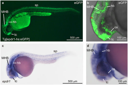
Establishment of Tg[epdr1-hs:eGFP]. (a,b) A heterozygous Tg[epdr1-hs:eGFP] embryo. eGFP expression was detected in the telencephalon (tc), the midbrain-hindbrain boundary (MHB), the hindbrain (hb) and spinal neurons (sp) at 30 hpf. The epdr1 expression in the optic stalk (os) was detected by confocal microscope (b). Autofluorescence of epidermis was detected (*). (c,d) Expression of epdr1 in the tc, the os, the MHB, the hb and the sp at 24 hpf. The expression of epdr1 was examined by whole-mount in situ hybridization using an anti-sense epdr1 RNA probe. |
Expression Data
| Genes: | |
|---|---|
| Fish: | |
| Anatomical Terms: | |
| Stage Range: | Prim-5 to Prim-15 |
Expression Detail
Antibody Labeling
Phenotype Data
Phenotype Detail
Acknowledgments
This image is the copyrighted work of the attributed author or publisher, and
ZFIN has permission only to display this image to its users.
Additional permissions should be obtained from the applicable author or publisher of the image.
Full text @ Sci. Rep.