
FIGURE
Fig. 4
- ID
- ZDB-FIG-170906-17
- Publication
- Magadum et al., 2017 - Live cell screening platform identifies PPARδ as a regulator of cardiomyocyte proliferation and cardiac repair
- Other Figures
- All Figure Page
- Back to All Figure Page
Fig. 4
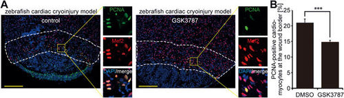
PPARδ activity is required for adult cardiomyocyte cell cycle activity after injury in zebrafish. (A) Representative images of cryoinjured zebrafish hearts (7 dpi) treated with DMSO (control) or 5 μM PPARδ inhibitor (GSK3787), stained for PCNA (green) and the cardiomyocyte marker Mef2 (red). Nuclei were visualized using DAPI (blue). Dashed lines highlight the wound border zone used for quantification. Scale bar = 150 μm. (B) Quantitative analysis of PCNA-positive cardiomyocytes (DMSO: n = 9 hearts; GSK3787: n = 10 hearts; ***P < 0.001). |
Expression Data
Expression Detail
Antibody Labeling
Phenotype Data
Phenotype Detail
Acknowledgments
This image is the copyrighted work of the attributed author or publisher, and
ZFIN has permission only to display this image to its users.
Additional permissions should be obtained from the applicable author or publisher of the image.
Full text @ Cell Res.