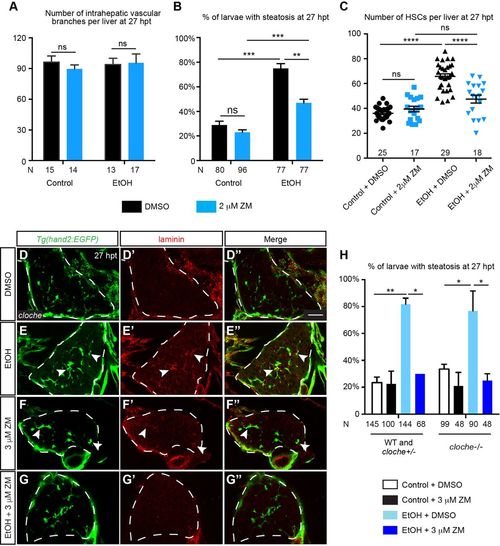
Ethanol-induced fibrogenesis and steatosis could be uncoupled from angiogenesis. (A) Numbers (mean±s.e.m.) of intrahepatic vascular branches per liver at 27 hpt in uninjured WT larvae treated with DMSO, uninjured larvae treated with 2 μM ZM, ethanol-exposed larvae treated with DMSO, and ethanol-exposed larvae treated with 2 μM ZM. (B) Percentages (mean±s.e.m.) of animals in different experimental groups with hepatic steatosis at 27 hpt based on Oil Red O staining. (C) Numbers (mean±s.e.m.) of HSCs per liver in different experimental groups at 27 hpt. (D-G″) Confocal single-plane images from the vibratome transverse sections showing that ZM treatment after acute ethanol exposure suppressed laminin deposition in cloche mutants. (D-G) HSCs marked by Tg(hand2:EGFP) expression; (D′-G′) laminin deposition; and (D″-G″) merged images of the two. Ventral views, anterior is to the top. Dashed line marks the liver. Scale bar: 30 μm. Ten larvae were examined in each experimental group. Arrowheads mark the HSCs that secreted laminin. (H) Percentages (mean±s.e.m.) of WT plus cloche heterozygotes, and homozygous cloche mutants treated with ethanol followed by ZM that exhibited hepatic steatosis at 27 hpt based on Oil Red O staining. Statistical significance in A,B,H was calculated by two-tailed Student's t-test, and in C one-way ANOVA and Tukey's post-hoc test. *P<0.05; **P<0.01; ***P<0.001; ****P<0.0001; ns, not significant.
|
