
FIGURE
Fig. S1
- ID
- ZDB-FIG-160818-40
- Publication
- Rochon et al., 2016 - Alk1 controls arterial endothelial cell migration in lumenized vessels
- Other Figures
- All Figure Page
- Back to All Figure Page
Fig. S1
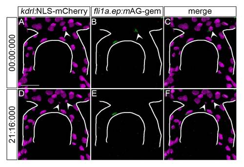
Tg(fli1a.ep:mAG-geminin)pt525 marks proliferating endothelial cells. 2D maximum projections of t = 0, ~36 hpf (A-C) and t = 21 min 16 sec (D-F) from confocal/two-photon timelapse imaging of a Tg(kdrl:NLS-mCherry)is4; Tg(fli1a.ep:mAG-geminin)pt525 embryo. Endothelial cell nuclei, magenta; endothelial S/G2/M-phase nuclei, green. Arrowheads, dividing cell (A-C) and progeny (D-F) transiently marked by mAG-geminin. The caudal division of the internal carotid artery (CaDI) is outlined in white. Frontal view, anterior up. Scale bar, 50 µm. Representative of N = 3 independent embryos. |
Expression Data
Expression Detail
Antibody Labeling
Phenotype Data
Phenotype Detail
Acknowledgments
This image is the copyrighted work of the attributed author or publisher, and
ZFIN has permission only to display this image to its users.
Additional permissions should be obtained from the applicable author or publisher of the image.
Full text @ Development