
Fig. 3
- ID
- ZDB-FIG-150513-14
- Publication
- Falcinelli et al., 2015 - Lactobacillus rhamnosus lowers zebrafish lipid content by changing gut microbiota and host transcription of genes involved in lipid metabolism
- Other Figures
- All Figure Page
- Back to All Figure Page
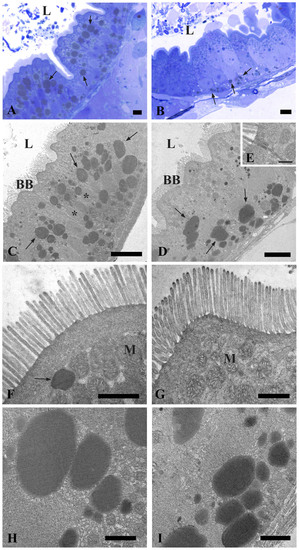
Transmission Electron Microscopy (TEM) shows the ultrastructure of the intestine in control and probiotic treated zebrafish. Thin section (80nm) of 6dpf zebrafish showing the epithelial layer arranged into broad, irregular folds (A). Intact epithelial barrier, lack of cell debris in the lumen and no signs of damage in larvae exposed to probiotic L. rhamnosus (B). Electron micrographs showing uniform, columnar, polarized epithelia, with an apical brush border in control (C) and probiotic treated groups (D). Higher magnification of junctional complexes (E). Microvilli on the apical surface and copious spherical mitochondria in the enterocyte cytoplasm of control (F) and treated larvae (G). Higher magnification of the lipid droplets in the enterocytes of control larvae (H) and in the probiotic treated intestine (I). BB: brush border; L: lumen; M: mitochondria; *: nucleus; arrows: lipid droplets. Scale bar: 5µm in (A, B); 10µm in (C, D); 500nm in (E), 1µm in (F, G); 2µm in (H, I). (See also Figure S3). |