
FIGURE
Fig. S3
- ID
- ZDB-FIG-150319-17
- Publication
- Takeuchi et al., 2015 - Establishment of Gal4 transgenic zebrafish lines for analysis of development of cerebellar neural circuitry
- Other Figures
- All Figure Page
- Back to All Figure Page
Fig. S3
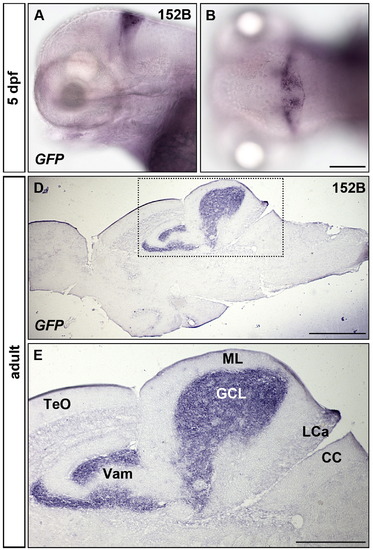
Localization of GFP transcripts in granule cells of gSA2AzGFF152B; UAS:GFP. (A, B) Expression of GFP at 5 dpf was detected by whole mount in situ hybridization. Lateral (A) and dorsal views (B) with anterior to the left. (D, E) Expression of GFP in the adult brain. In situ hybridization of sagittal sections. Anterior to the left. (E) High magnification view of a box in D. Scale bars: 200 µm in B (applied to A, B); 50 µm in D; 25 µm in E. Note that GFP transcripts are detected only in the somata of granule cells (D, E). |
Expression Data
Expression Detail
Antibody Labeling
Phenotype Data
Phenotype Detail
Acknowledgments
This image is the copyrighted work of the attributed author or publisher, and
ZFIN has permission only to display this image to its users.
Additional permissions should be obtained from the applicable author or publisher of the image.
Reprinted from Developmental Biology, 397(1), Takeuchi, M., Matsuda, K., Yamaguchi, S., Asakawa, K., Miyasaka, N., Lal, P., Yoshihara, Y., Koga, A., Kawakami, K., Shimizu, T., Hibi, M., Establishment of Gal4 transgenic zebrafish lines for analysis of development of cerebellar neural circuitry, 1-17, Copyright (2015) with permission from Elsevier. Full text @ Dev. Biol.