
Fig. 1
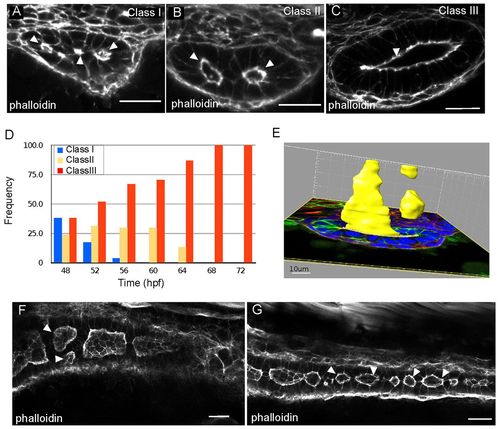
Lumens enlarge and fuse during single lumen formation in the zebrafish gut. (A-C) Confocal images of cross sections of wild-type embryos exhibiting class I (A), class II (B) and class III (C) lumens, stained with phalloidin. Arrowheads indicate the lumens. (D) Quantification of lumen phenotypes between 48 and 72 hpf: 48 hpf n=21, 52 hpf n=29, 56 hpf n=27, 60 hpf n=27, 64 hpf n=30, 68 hpf n=21, 72 hpf n=26. (E) Space-fill projection from a 200 7μm confocal stack of an intestine section at the resolution stage. Yellow, lumen; green, GFP-CaaX; blue, DAPI. (F) Confocal whole-mount image of the anterior gut at 58 hpf stained with phalloidin (red). Arrowheads point to adjacent unfused lumens. (G) Confocal whole-mount image of the posterior gut at 58 hpf stained with phalloidin (red). Arrowheads point to lumens. Scale bars: 20 μm in A-C; 10 μm in E; 20 μm in F,G. |