
FIGURE
Fig. S7
- ID
- ZDB-FIG-140422-54
- Publication
- Holly et al., 2014 - Sfrp1a and Sfrp5 function as positive regulators of Wnt and BMP signaling during early retinal development
- Other Figures
- All Figure Page
- Back to All Figure Page
Fig. S7
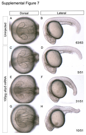
Morphological assessment of zebrafish embryos injected with 100 pg of sfrp5 mRNA. One-cell stage embryos were injected with a high dose (100 pg) of sfrp5 mRNA. Embryos were grown to 22 hpf and analyzed for gross morphological defects. Uninjected controls display no aberrations in either eye or alterations to axis specification (A-B, n=63/63). Of the embryos injected with a high dose of sfrp5 mRNA, 5/51 display no overt phenotype (C-D), 31/51 display subtly elongated eyes and very mild dorsalization (E-F), and 10/51 display extensive elongation of eyes as well as moderate dorsalization (G-H). |
Expression Data
Expression Detail
Antibody Labeling
Phenotype Data
Phenotype Detail
Acknowledgments
This image is the copyrighted work of the attributed author or publisher, and
ZFIN has permission only to display this image to its users.
Additional permissions should be obtained from the applicable author or publisher of the image.
Reprinted from Developmental Biology, 388(2), Holly, V.L., Widen, S.A., Famulski, J.K., and Waskiewicz, A.J., Sfrp1a and Sfrp5 function as positive regulators of Wnt and BMP signaling during early retinal development, 192-204, Copyright (2014) with permission from Elsevier. Full text @ Dev. Biol.