
FIGURE
Fig. 1
- ID
- ZDB-FIG-131009-7
- Publication
- Hisano et al., 2013 - Functional cooperation of spns2 and fibronectin in cardiac and lower jaw development
- Other Figures
- All Figure Page
- Back to All Figure Page
Fig. 1
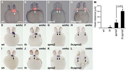
Cardiac morphology.Heart positions are indicated by the arrowheads. (A–D) Cardiac morphology visualized by mRFP expression derived from Tg(cmlc2:mRFP). All images show ventral views at 28hpf. (E–L) Whole-mount in situ hybridization with amhc and vmhc RNA probes. All images show ventral views at 30hpf except for panel L (dorsal view). Genotyping was performed by genomic sequencing after taking pictures, wt (A,E,I), fn mutant (B,F,J), spns2 mutant (C,G,K) and fn;spns2 double mutant (D,H,L). Scale bars: 200μm. (M) Average distances between two hearts from multiple experiments; error bars represent standard deviations. |
Expression Data
| Genes: | |
|---|---|
| Fish: | |
| Anatomical Term: | |
| Stage Range: | Prim-5 to Prim-15 |
Expression Detail
Antibody Labeling
Phenotype Data
| Fish: | |
|---|---|
| Observed In: | |
| Stage Range: | Prim-5 to Prim-15 |
Phenotype Detail
Acknowledgments
This image is the copyrighted work of the attributed author or publisher, and
ZFIN has permission only to display this image to its users.
Additional permissions should be obtained from the applicable author or publisher of the image.
Full text @ Biol. Open