
FIGURE
Fig. S5
Fig. S5
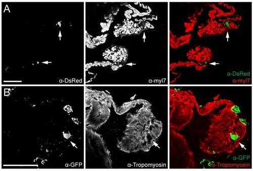
Isl1BAC reporter activity in adult heart. Confocal images of Tg(isl1BAC:GalFF; UAS:RFP; myl7:eGFP) after immunolabeling with anti-RFP and anti-GFP antibodies (A), or Tg(isl1BAC:GalFF; UAS:GFP) after immunolabeling with anti-GFP and antitropomyosin antibodies (B). Isl1 expressing cells (indicated with arrows) are located at the base of the venous valves and contain much lower levels of myosin light chain or tropomyosin compared to surrounding myocardial cells. Axonal Isl1/GFP+structures are visible at the outer surface of the myocardium. Scale bars represent 50 μm |
Expression Data
Expression Detail
Antibody Labeling
Phenotype Data
Phenotype Detail
Acknowledgments
This image is the copyrighted work of the attributed author or publisher, and
ZFIN has permission only to display this image to its users.
Additional permissions should be obtained from the applicable author or publisher of the image.
Full text @ PLoS One