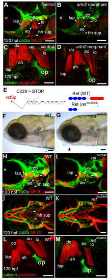
Loss of Ret or Artn2 function results in specific opercular muscle defects. (A-D,H-M) Immunolabelling of cartilage, bone (green) and muscles (red) of uninjected controls (A,C), artn2 morphants (B,D), wild type (WT; H,J,L) and rethu2846 mutants (I,K,M) at 120 hours post-fertilisation (hpf). Specific reductions of the do, ao, lo and hh sup muscles occur in artn2 morphants and rethu2846 mutants, but the lap and ah muscles are unaffected. (E-G) rethu2846 mutants have a tightly closed mouth (G, arrowhead) relative to WT (F) and have a point mutation in codon 229 (T>A) of exon 4 in the ret gene that changes a cysteine to a stop codon in the coding sequence (E). This results in a truncated protein lacking the tyrosine kinase domain (red), the transmembrane region (green) and part of the extracellular domain (blue). A morpholino directed to the exon 1 splice donor site (retSp) causes aberrant splicing of ret. ah, adductor hyoideous; ao, adductor operculi; do, dilator operculi; e, eye; hh sup, hyohyoideus superiores; lap, levator arcus palatini; lo, levator operculi. Scale bars: 100 μm in A,B,H-K; 50 μm in C,D,L,M.
|
