
FIGURE
Fig. 3
- ID
- ZDB-FIG-100903-43
- Publication
- Wang et al., 2010 - Moesin1 and Ve-cadherin are required in endothelial cells during in vivo tubulogenesis
- Other Figures
- All Figure Page
- Back to All Figure Page
Fig. 3
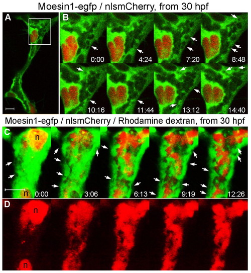
Vacuoles fuse with luminal membranes during initial lumen expansion. (A) An ISV from a living Tg(flk1:moesin1-egfp)/Tg(flk1:nlsmCherry) zebrafish embryo with the box indicating the area shown in B. (B) Time-lapse confocal images showing formation of primary lumen in an ISV with the integration of vacuoles (arrows) from 30 hpf. (C,D) A dorsal ISV from a living Tg(flk1:moesin1-egfp)/Tg(flk1:nlsmCherry) embryo injected intravenously with labeled dextran (D) showing the intracellular vacuoles without labeled dextran (C, arrows). The time is in minutes:seconds. n, nucleus. |
Expression Data
| Genes: | |
|---|---|
| Fish: | |
| Anatomical Terms: | |
| Stage: | Prim-15 |
Expression Detail
Antibody Labeling
Phenotype Data
Phenotype Detail
Acknowledgments
This image is the copyrighted work of the attributed author or publisher, and
ZFIN has permission only to display this image to its users.
Additional permissions should be obtained from the applicable author or publisher of the image.
Full text @ Development