
Fig. 4
- ID
- ZDB-FIG-100209-5
- Publication
- Jung et al., 2010 - Visualization of myelination in GFP-transgenic zebrafish
- Other Figures
- All Figure Page
- Back to All Figure Page
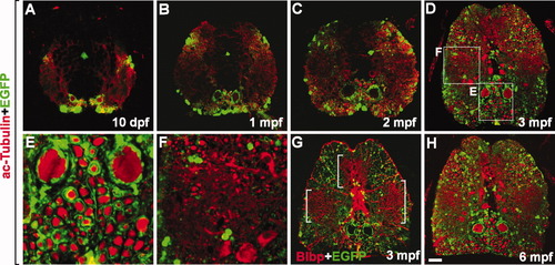
Axon myelination occurs continuously in the spinal cord of postembryonic zebrafish. All images are transverse sections of the spinal cord of Tg(mbp:egfp) zebrafish, dorsal side up. Stages are indicated on each panel. A-F,H: Labeling with anti-acetylated tubulin antibody to mark axons. E,F: High magnification images of boxed areas in D. G: Labeling with anti-Blbp antibody to mark radial glia. Bracketed areas indicate clusters of highly branched radial glial processes. Scale bars = 25 μm in A, 50 μm in B, 80 μm in C, 100 μm in D,G,H, 25 μm in E,F. |