
FIGURE
Fig. 2
Fig. 2
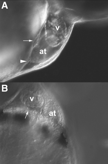
Proepicardial organ lies in apposition to the myocardium of the embryonic heart. (A) A lateral view of the zebrafish heart at about 50 hpf with clusters of spherical cells seen using differential interference contrast optics nestled between the atrium and the yolk (arrow) and adjacent to the sinus venosus (arrowhead). Anterior is to the right. (B) Ventral view of the heart (50 hpf) with a cluster of cells attached to the outer ventricular wall (arrow). Anterior is at the top of the panel. at: atrium, v: ventricle. Movies of these embryos can be found in the supplemental information section. |
Expression Data
Expression Detail
Antibody Labeling
Phenotype Data
Phenotype Detail
Acknowledgments
This image is the copyrighted work of the attributed author or publisher, and
ZFIN has permission only to display this image to its users.
Additional permissions should be obtained from the applicable author or publisher of the image.
Reprinted from Developmental Biology, 315(1), Serluca, F.C., Development of the proepicardial organ in the zebrafish, 18-27, Copyright (2008) with permission from Elsevier. Full text @ Dev. Biol.