
FIGURE
Fig. S6
- ID
- ZDB-FIG-080108-11
- Publication
- Murayama et al., 2006 - Tracing Hematopoietic Precursor Migration to Successive Hematopoietic Organs during Zebrafish Development
- Other Figures
- All Figure Page
- Back to All Figure Page
Fig. S6
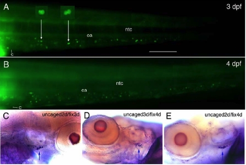
Dynamics of Cell Seeding from AGM to CHT and Thymus (A,B) Fluorescent cells at 3 and 4 dpf in the ventral tail (CHT) of a live embryo in which fluorescein-dextran was laser-uncaged along the entire DP joint at 2 dpf (end of uncaged area visible to the left). In (A), two putative pairs of sister cells are shown magnified (arrows). Bar, 100 μm. (C-E) Immunodetection of labelled cells in the right (C) or left (D,E) thymus (arrows) following uncaging in the DP joint and later fixation at the indicated stages. |
Expression Data
Expression Detail
Antibody Labeling
Phenotype Data
Phenotype Detail
Acknowledgments
This image is the copyrighted work of the attributed author or publisher, and
ZFIN has permission only to display this image to its users.
Additional permissions should be obtained from the applicable author or publisher of the image.
Reprinted from Immunity, 25(6), Murayama, E., Kissa, K., Zapata, A., Mordelet, E., Briolat, V., Lin, H.F., Handin, R.I., and Herbomel, P., Tracing Hematopoietic Precursor Migration to Successive Hematopoietic Organs during Zebrafish Development, 963-975, Copyright (2006) with permission from Elsevier. Full text @ Immunity