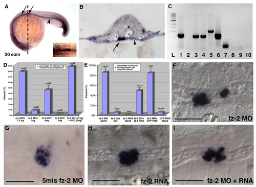
Migration defects in Fz-2 morphant embryos can be rescued by synthetic fz-2 mRNA. (A) Double in situ hybridization with fz-2 and insulin at 20 somite stage of development. Arrow, insulin; arrowhead, fz-2 expression in the endoderm; dotted line, approximate position of the section in (B). (B) A section of double in situ hybridization with fz-2 and insulin. Fz-2 is expressed more strongly on the surface of mesoderm and entire endoderm. Arrow, insulin; arrowhead, fz-2 expression in the endoderm; a, arteries; asterisk, neural tube; d, pronephric duct. (C) RT-PCR using cDNA made from sorted cells of transgenic insulin:GFP zebrafish embryos. L: ladder; lanes 1-5: GFP-negative cells; lanes 6-10: GFP-positive cells; lanes 1, 6: EF1α lanes 2, 7: insulin; lanes 3, 8: fz-2 primer set #1; lanes 4, 9: fz-2 primer set #2; lanes 5, 10: wnt-5. (D) High-dose injection of either fz-2 MO1 or MO2 resulted in scattered insulin expression, whereas low dose injection of either MO caused such defects in less than 10% of embryos. Co-injection of low dose fz-2 MO1 and MO2 resulted in synergistic increase of percentage of embryos with scattered insulin expression. (E) 80% of fz-2 MO-injected embryos displayed scattered insulin expression. Co-injection of fz-2 MO and fz-2 RNA reduced the percentage of embryos with abnormal insulin expression down to 45%. (F-I) In situ hybridization with insulin at 24 hpf stage, anterior is to the left, (F) fz-2 MO1-injected embryo, (G) fz-2 mismatch MO-injected embryo, (H) fz-2 RNA-injected embryo, (I) fz-2 MO- and fz-2 RNA-co-injected embryo. Notice the compact islet in this embryo that displays an undulated notochord.
|
