ZFIN is incorporating published figure images and captions as part of an ongoing project. Figures from some publications have not yet been curated, or are not available for display because of copyright restrictions.

PHENOTYPE:
Fish:
Knockdown Reagents:
Observed In:
Stage: Day 5

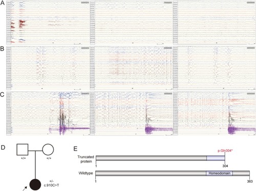
The clinical data and
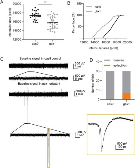
 GBX1 variant in the patient. A–C. Abnormal EEG. (A) Normal background rhythm with 8–10 Hz α waves in occipitals during wakefulness with eyes closed. High‐amplitude slow wave bursts in the right frontal region and sharp‐slow wave bursts in both occipitals. (B) During sleep, generalized spikes, spike‐slow waves, and polyspike bursts, especially in the left frontal region, with scattered bursts in the right frontal, occipital, and posterior temporal regions. (C) Focal seizures originating from the right frontal region, evolving from slow waves to fast‐wave activity. (D) The pedigree for the family. The patient was highlighted with an arrow with the filled symbol. (E) The schematic diagram for wildtype and p.Gln304* GBX1 protein.

Morphology and electrophysiology of gbx1‐cas9 zebrafish. (A, B) The interocular area in control cas9 and gbx1‐cas9 groups. (C) Electrophysiological testing in two groups. (D) The distribution of zebrafish with baseline and epileptiform signals in the cas9 control group and the gbx1‐cas9 group. ***p < 0.001.

Acknowledgments
This image is the copyrighted work of the attributed author or publisher, and ZFIN has permission only to display this image to its users. Additional permissions should be obtained from the applicable author or publisher of the image. Full text @ Mol Genet Genomic Med