FIGURE SUMMARY
Title

Biallelic variants in CCN2 underlie an autosomal recessive kyphomelic dysplasia

Authors
Singh, S., Danda, S., Sharma, N., Shah, H., Madhuri, V., Mir, T.A., Padala, N.Z., Medishetti, R., Ekbote, A., Bhavani, G.S., Sevilimedu, A., Girisha, K.M.
Source
Full text @ Eur. J. Hum. Genet.

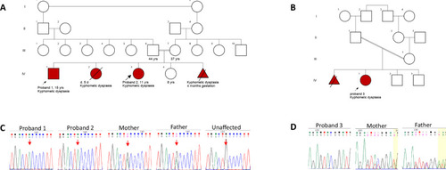
Pedigrees and Sanger chromatograms.

A Pedigree of the family 1 and B family 2 depicting consanguinity and affected probands with kyphomelic dysplasia. C Sanger chromatograms in family 1 show the variant c.448G>A in homozygous state in both affected siblings, heterozygous state in their parents and absent in their unaffected sibling. D Chromatograms of family 2 confirm the variant c.779_786delG>A in homozygous state in proband 3 and heterozygous state in her parents.

Kyphomelic femora in the participants.

Radiographs of pelvis [proband 1 at 15 years (A), proband 2 at 11 years (B) and proband 3 at 3 years of age (C)] show short and broad pelvis, coxa vara and reduced hip joint space in all. Kyphotic femora are seen in all of them but is more prominent (arrows) in the younger proband 3 (C).

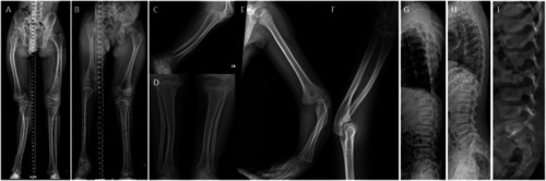
Bowing of long bones in the affected individuals.

Radiographs of limbs of probands: proband 1 at 15 years (
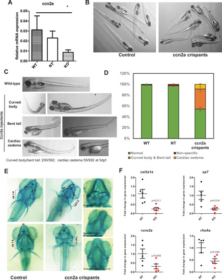
A), proband 2 at 11 years (B) and proband 3 at 3 years of age (CF) show bowing of long bones. Bowing of femur (A and B), bowing of the tibia and fibula (A, C, D) can be observed. Irregular epiphyses and metaphyses along with flaring of metaphyses at the knee joint can be noted (D). Variable platyspondyly can be observed in all (GI).

Phenotypes in the ccn2a FO knockout zebrafish.

A Relative expression levels of the ccn2a mRNA in the controls (NT) and injectants (crispants). B Bright field image of a group of ccn2a crispants and matched control injectants from the same experiment to illustrate the extent of physical phenotypes. C Representative images of observed phenotypes as labeled, among the ccn2a crispants. D Quantification of the number of larvae showing the indicated phenotypes in each group. E Alcian blue stained images of wildtype and representative examples of ccn2a crispants, showing cartilage deformities. Cartilage elements labeled: m: Meckel’s cartilage; ch: ceratohyal; cb: ceratobranchial pairs. F Fold change in mRNA expression of select markers such as col2a1a (chondrocyte), sp7 (osteoblast), runx2a (early osteoblast) and rho1a (palatogenesis) in the ccn2a crispants as compared to the controls at 5dpf. Results from at least 5 independent experiments (pooled larvae, 30–50/experiment) are quantified.

Acknowledgments
This image is the copyrighted work of the attributed author or publisher, and ZFIN has permission only to display this image to its users. Additional permissions should be obtained from the applicable author or publisher of the image. Full text @ Eur. J. Hum. Genet.