
PUBLICATION
Endothelial Cell Self-fusion during Vascular Pruning
- Authors
- Lenard, A., Daetwyler, S., Betz, C., Ellertsdottir, E., Belting, H.G., Huisken, J., Affolter, M.
- ID
- ZDB-PUB-150418-3
- Date
- 2015
- Source
- PLoS Biology 13(4): e1002126 (Journal)
- Registered Authors
- Affolter, Markus, Belting, Heinz-Georg Paul (Henry), Ellertsdottir, Elin, Huisken, Jan, Lenard, Anna
- Keywords
- SIV, Embryos, Blood vessels, Endothelial cells, Cell fusion, Blood flow, Cell membranes, Zebrafish
- MeSH Terms
-
- Zebrafish/embryology
- Animals, Genetically Modified
- Neovascularization, Physiologic
- Animals
- Cell Fusion*
- PubMed
- 25884426 Full text @ PLoS Biol.
Abstract
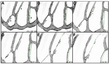
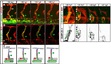
During embryonic development, vascular networks remodel to meet the increasing demand of growing tissues for oxygen and nutrients. This is achieved by the pruning of redundant blood vessel segments, which then allows more efficient blood flow patterns. Because of the lack of an in vivo system suitable for high-resolution live imaging, the dynamics of the pruning process have not been described in detail. Here, we present the subintestinal vein (SIV) plexus of the zebrafish embryo as a novel model to study pruning at the cellular level. We show that blood vessel regression is a coordinated process of cell rearrangements involving lumen collapse and cell-cell contact resolution. Interestingly, the cellular rearrangements during pruning resemble endothelial cell behavior during vessel fusion in a reversed order. In pruning segments, endothelial cells first migrate toward opposing sides where they join the parental vascular branches, thus remodeling the multicellular segment into a unicellular connection. Often, the lumen is maintained throughout this process, and transient unicellular tubes form through cell self-fusion. In a second step, the unicellular connection is resolved unilaterally, and the pruning cell rejoins the opposing branch. Thus, we show for the first time that various cellular activities are coordinated to achieve blood vessel pruning and define two different morphogenetic pathways, which are selected by the flow environment.
Genes / Markers
Figure Gallery (7 images)


Expression
Phenotype
Mutations / Transgenics
Human Disease / Model
Sequence Targeting Reagents
Fish
Orthology
Engineered Foreign Genes
Mapping