
Fig. 5
- ID
- ZDB-IMAGE-260530-143
- Publication
- Tsai et al., 2026 - Zebrafish gon4la mutants recapitulate human GON4L-related growth disorders and reveal novel metabolic organs abnormalities
- All Figures
- Figures for Tsai et al., 2026
|
Fig. 5
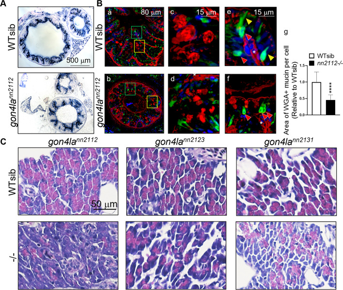
Impaired intestinal epithelial differentiation and pancreatic exocrine defects in gon4la mutants. (A) Alkaline phosphatase (AP) staining for enterocyte differentiation. (B) Immunofluorescent staining for goblet cells and enteroendocrine cells. (a, b) Cross section of posterior intestine at lower magnification. (c, d) Magnified views (from yellow squares) of goblet cells stained with WGA (red signals). (e, f) Magnified views (from green squares) of enteroendocrine cells (green signals, 2F11 antibody). Yellow arrowheads indicate matured enteroendocrine cells exhibited polarized morphology. Red arrowheads indicate less matured enteroendocrine cells. (g) WGA-positive mucin area within the goblet cell theca, shown as a ratio relative to WT siblings. Measurements were taken from n = 73 thecae from 4 WT sibling fish and n = 49 thecae from 4 gon4lann2112 homozygous mutant fish. ****P < 0.0001. Quantification of the WGA-positive mucin area (theca size) relative to WT siblings shows significantly reduced secretory capacity in mutants. (C) H&E staining of pancreatic exocrine in WT and gon4la mutant fish. H&E staining of exocrine pancreas from adult WT and mutant fish at 3–4 mpf. Acinar cells in all mutant lines show marked reduction of zymogen granules.