IMAGE

Fig. 7

ID
ZDB-IMAGE-250512-61
Antibodies
Source
Figures for Fargher et al., 2025
Image
Figure Caption

Fig. 7 acmsd-/- zebrafish retain normal numbers and activation of microglia.

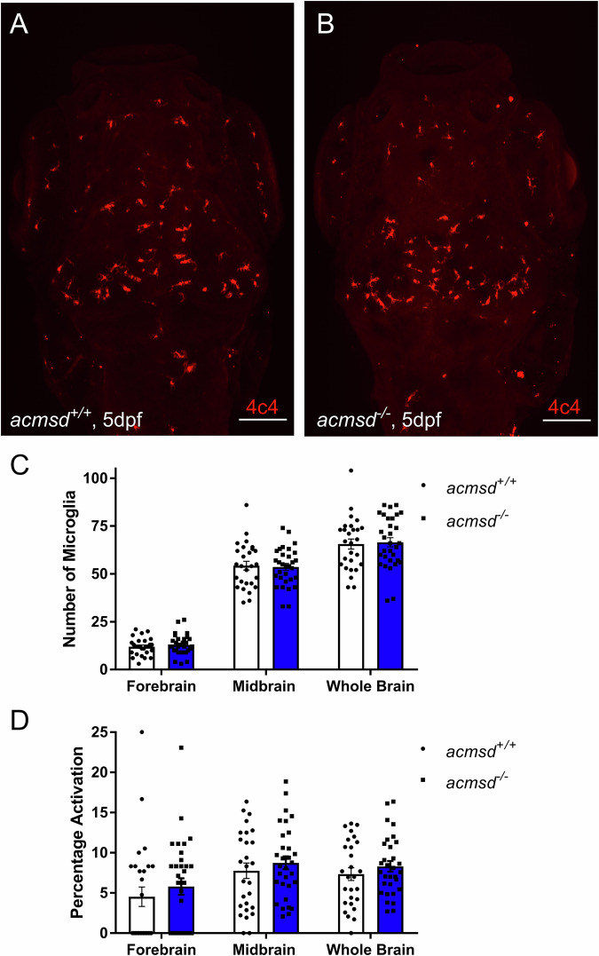
Representative images from wholemount immunohistochemistry against 4c4 in acmsd+/+ (A) and acmsd-/- (B) siblings at 5dpf (scale bar = 100 µm). C No difference was found in the number of 4c4+ cells between acmsd+/+ and acmsd-/- larvae in the forebrain, midbrain, or these combined (whole brain) (p = 0.9827 for forebrain, p = 0.9925 for midbrain, p = 0.9836 for whole brain). D No difference in microglial activation, represented as the percentage of amoeboid cells out of the total 4c4+ cell count, was identified between genotypes (p = 0.6983 for forebrain, p = 0.8307 for midbrain, p = 0.8441 for whole brain). n = 31 acmsd-/-, 27 acmsd+/+, from three biological replicates. Statistics analysed by two-way ANOVA with post hoc Sidak’s multiple comparisons test.

Figure Data
Acknowledgments
This image is the copyrighted work of the attributed author or publisher, and ZFIN has permission only to display this image to its users. Additional permissions should be obtained from the applicable author or publisher of the image. Full text @ NPJ Parkinsons Dis