IMAGE

Fig. 6

ID
ZDB-IMAGE-250512-60
Genes
Source
Figures for Fargher et al., 2025
Image
Figure Caption

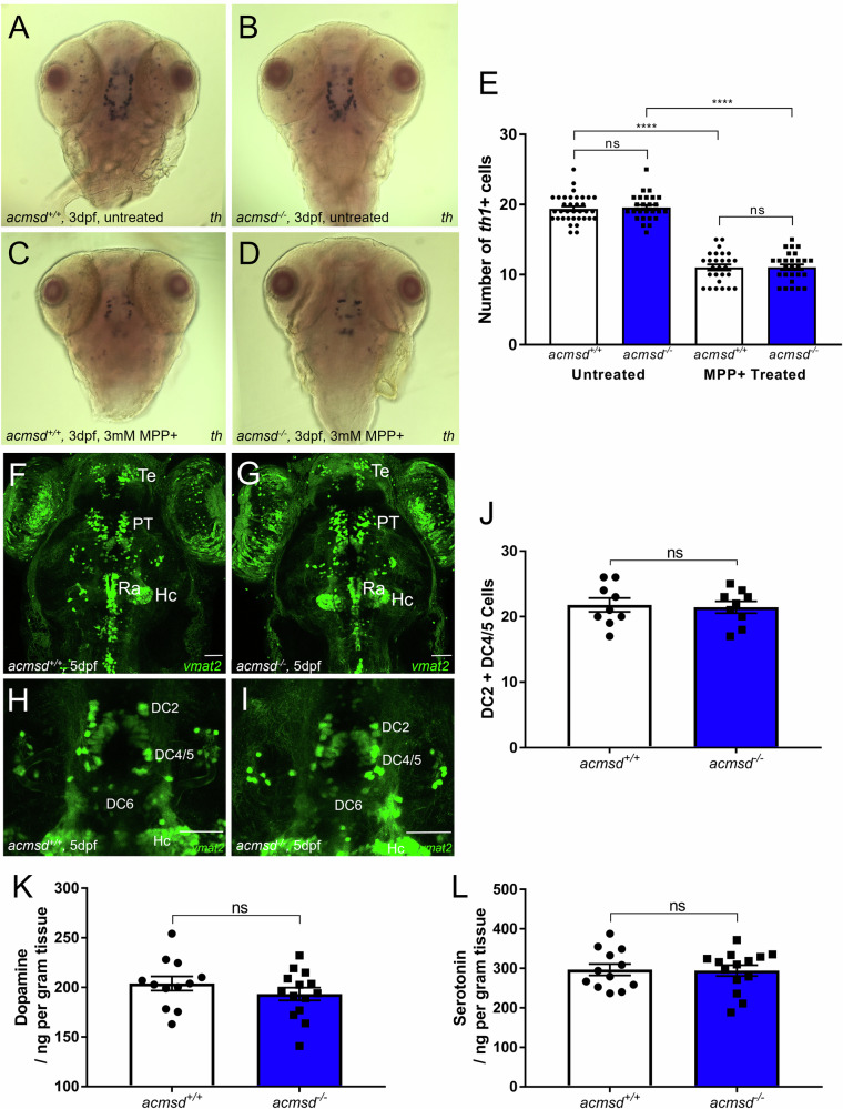
Fig. 6 acmsd-/- zebrafish retain normal dopaminergic neuron numbers and susceptibility to MPP +.

AD In situ hybridisation against th1 showed no change in staining pattern between acmsd+/+ and acmsd-/- zebrafish either before or after MPP+ exposure. E There was no difference in th1+ neuron numbers in the ventral diencephalon between acmsd+/+ and acmsd-/- zebrafish (p = 0.9866). MPP+ exposure resulted in significant reductions in th1+ cells irrespective of genotype (43.1% in acmsd+/+; 43.4% in acmsd-/-, p < 0.0001). Total cell counts between groups following exposure remained indistinguishable (p = 0.9999). n = 26–35 per group, from three biological replicates. Statistics from a two-way ANOVA with Tukey’s multiple comparisons post-hoc test. Monoaminergic neurons were visualised using the ETvmat2:GFP transgenic line at 5dpf in the full brain (F, G dorsal view) and in the DC neuronal groups (H, I) in both acmsd+/+ and acmsd-/- zebrafish. Scale bar = 50 µm. DC diencephalic neurons, Hc caudal hypothalamus, PT pretectal neural cluster, Ra raphe nucleus, Te telencephalic neurons. J No difference in the number of neuronal cell bodies in DC2 and DC4/5 was seen between acmsd+/+ and acmsd-/- zebrafish (p = 0.8126, unpaired two-tailed t test). n = 9 per genotype, from three biological replicates.No difference in the concentrations of dopamine (K, p = 0.2808) or serotonin (L, p = 0.9079) were identified in whole 11mpf brains. Data analysed using an unpaired two-tailed t test. n = 14 acmsd-/-; 12 acmsd+/+, 1 brain per replicate.

Figure Data
Acknowledgments
This image is the copyrighted work of the attributed author or publisher, and ZFIN has permission only to display this image to its users. Additional permissions should be obtained from the applicable author or publisher of the image. Full text @ NPJ Parkinsons Dis